FIGURE
Fig. S3
Fig. S3
|
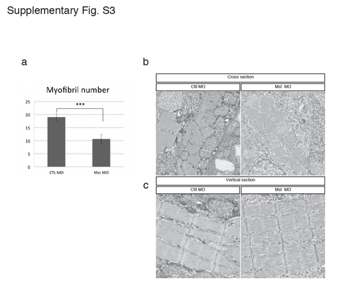
Electron micrograph of the slow muscle structure in the mysterin morphant. (a) Quantification of myofibril numbers in 5 μm2 square of cross-sectionnal electron micrograph. Control and mysterin MOs show 19.0 ± 1.4 (n=12) and 10.6 ± 1.6 (n=11) myofibrils in 5 μm2 square, respectively. ***P < 0.001. Cross-section (b) and vertical section (c) of transgenic animals (gSA2AzGFF598A) that express Gal4FF in fast muscle. Slow muscle is identified by its distribution and the thickness of the Z-disk. Treatment with a low dose of the MO targeting mysterin-α does not affect slow muscle structures. |
Expression Data
Expression Detail
Antibody Labeling
Phenotype Data
Phenotype Detail
Acknowledgments
This image is the copyrighted work of the attributed author or publisher, and
ZFIN has permission only to display this image to its users.
Additional permissions should be obtained from the applicable author or publisher of the image.
Full text @ Sci. Rep.

