Fig. S2
- ID
- ZDB-FIG-170921-59
- Publication
- Gallagher et al., 2017 - Pnrc2 regulates 3'UTR-mediated decay of segmentation clock-associated transcripts during zebrafish segmentation
- Other Figures
- All Figure Page
- Back to All Figure Page
|
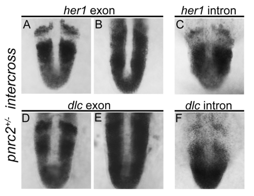
her1 and dlc transcripts accumulate post-transcriptionally in pnrc2oz22mutants. Exonic in situ probes reveal that segmentation clock-associated her1 and dlc transcripts are misexpressed in the expected one-quarter of embryos in a pnrc2oz22 intercross, n=13/57 (X2=0.1, p=0.7) and n=8/30 (X2=0.04, p=0.83), respectively (A, B and D, E). Intronic in situ probes, however, reveal no differences in expression among embryos from the same clutch, n=30/30 (X2=10.0, p=0.0016) and n=45/45 (X2=15.0, p=0.0001), respectively (C, F). These results are consistent with previous observations in torb644 mutants using intronic and exonic in situ probes that distinguish nascent from processed transcripts (Dill et al, 2005). |
Reprinted from Developmental Biology, 429(1), Gallagher, T.L., Tietz, K.T., Morrow, Z.T., McCammon, J.M., Goldrich, M.L., Derr, N.L., Amacher, S.L., Pnrc2 regulates 3'UTR-mediated decay of segmentation clock-associated transcripts during zebrafish segmentation, 225-239, Copyright (2017) with permission from Elsevier. Full text @ Dev. Biol.

