
FIGURE
Fig. 4
- ID
- ZDB-FIG-170103-30
- Publication
- Montalbano et al., 2016 - Retinoic acid catabolizing enzyme CYP26C1 is a genetic modifier in SHOX deficiency
- Other Figures
- All Figure Page
- Back to All Figure Page
Fig. 4
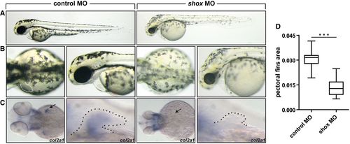
Pattern of defects in zebrafish embryos injected with anti‐shox morpholino. Wild‐type embryos injected with control MO or with shox MO. A.Lateral views of the embryos at 55 hours post‐fertilization (hpf). B. Dorsal view and magnification on the lateral view of the embryos. Dotted line, pectoral fins. shox morphants show smaller fins compared to controls (n = 30 embryos). C. Expression of col2a1 at 55 hpf was examined by in situ hybridization in wild‐type embryos injected with control MO and with shox MO. Arrow and dotted line indicate the pectoral fin. D. Pectoral fin area was measured by ImageJ (n = 30 embryos). |
Expression Data
| Gene: | |
|---|---|
| Fish: | |
| Knockdown Reagent: | |
| Anatomical Term: | |
| Stage: | Long-pec |
Expression Detail
Antibody Labeling
Phenotype Data
| Fish: | |
|---|---|
| Knockdown Reagent: | |
| Observed In: | |
| Stage: | Long-pec |
Phenotype Detail
Acknowledgments
This image is the copyrighted work of the attributed author or publisher, and
ZFIN has permission only to display this image to its users.
Additional permissions should be obtained from the applicable author or publisher of the image.
Full text @ EMBO Mol. Med.