
|
Fig. 4
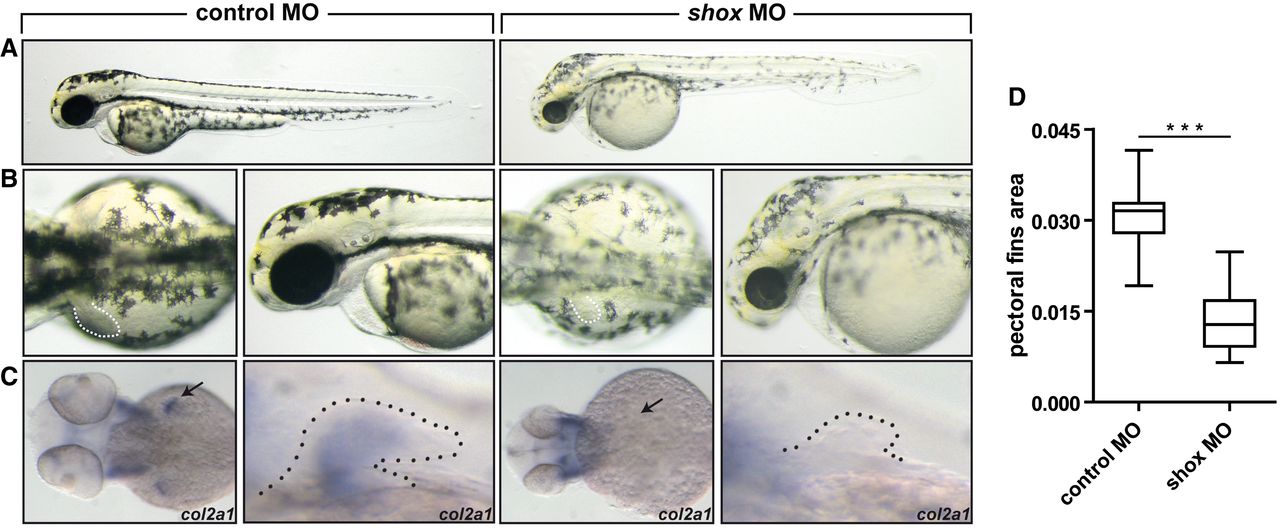
Pattern of defects in zebrafish embryos injected with anti‐shox morpholino.
Wild‐type embryos injected with control MO or with shox MO.
A.Lateral views of the embryos at 55 hours post‐fertilization (hpf).
B. Dorsal view and magnification on the lateral view of the embryos. Dotted line, pectoral fins. shox morphants show smaller fins compared to controls (n = 30 embryos).
C. Expression of col2a1 at 55 hpf was examined by in situ hybridization in wild‐type embryos injected with control MO and with shox MO. Arrow and dotted line indicate the pectoral fin.
D. Pectoral fin area was measured by ImageJ (n = 30 embryos).