
Fig. S8
- ID
- ZDB-FIG-150922-12
- Publication
- Casari et al., 2014 - A Smad3 transgenic reporter reveals TGF-beta control of zebrafish spinal cord development
- Other Figures
- All Figure Page
- Back to All Figure Page
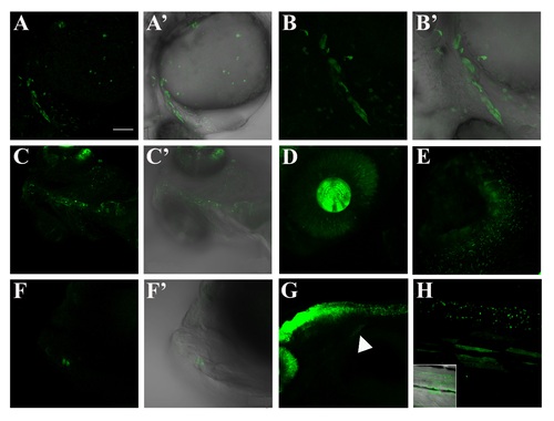
Confocal images of GFP-positive regions external to CNS during the first month of development. A-A′, confocal lateral view of 26 hpf embryo (Z-stack). Reporter gene is activated in the cardiac mesoderm and around the outflow tract. Spare cells are also visible in the yolk ball. B-B′, zoomed heart region of a 26 hpf embryo (Z-stack). C-C′, ventral view of heart and dorsal aorta of a 4 dpf larva (Z-stack). GFP expression in this area is no longer visible beyond this stage. D, zoomed view of the eye of a 48 hpf larva (Z-stack). GFP-positive cells are visible both in retina and lens. E, zoomed view of an olfactory pit of a 6 dpf larva (Z-stack). F-F′, magnified image of the jaw of a 5 dpf larva (Z-stack). Few cells are visible in this area during the early larval development; G, lateral image of a 48 hpf larva: hindbrain, spinal cord and fin bud (white arrowhead). H, zoomed lateral view of the trunk region of a 40 dpf larva, with the correspondent bright field. Reporter expression is still present in the neural tube. Some fibers of the median musculature seem to activate Smad3-mediated signaling during this developmental stage. Scale bar is 100 µm in A-A′, C-C′, F-G; 50 µm in B-B′, H; 20 µm in D; 10 µm in E. |
| Gene: | |
|---|---|
| Fish: | |
| Anatomical Terms: | |
| Stage Range: | Prim-5 to Days 30-44 |
Reprinted from Developmental Biology, 396(1), Casari, A., Schiavone, M., Facchinello, N., Vettori, A., Meyer, D., Tiso, N., Moro, E., Argenton, F., A Smad3 transgenic reporter reveals TGF-beta control of zebrafish spinal cord development, 81-93, Copyright (2014) with permission from Elsevier. Full text @ Dev. Biol.