
Fig. 1
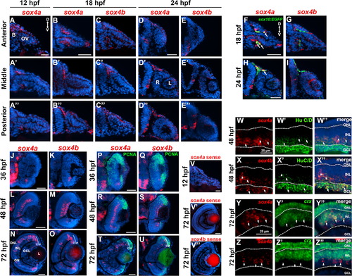
Expression of sox4a and sox4b during ocular development. (A–A′′) Fluorescent in situ hybridization (FISH) for sox4a performed on transverse cryosections at 12 hpf. (B–D′′) FISH for sox4a (B–B′′ and D–D′′) or sox4b (C–C′′ and E–E′′) performed on transverse cryosections taken at the level of the anterior, middle, and posterior optic vesicle at 18 or 24 hpf. (F–I) FISH with sox4a and sox4b probes performed on sections from sox10:EGFP transgenic embryos at 18 and 24 hpf. (J–O) FISH for sox4a or sox4b performed on transverse retinal cryosections at 36, 48, and 72 hpf. (P–U) FISH combined with immunohistochemistry for the proliferation marker PCNA at 24, 48, or 72 hpf. (V–V′′) Control FISH with a sense probe for sox4a and sox4b at 12 and 72 hpf. (W–X′′) FISH combined with immunohistochemistry for HuC/D, which labels ganglion and amacrine cells, at 48 hpf. (Y–Z′′) Two-color FISH with sox4a/b and crx probes performed at 72 hpf. Arrows indicate co-localization, arrowheads indicate sox4a/b+ cells that did not co-localize with the indicated marker. All scale bars in A-V equal 50 µm. D, dorsal; V, ventral; B, brain; OV, optic vesicle; L, lens; R, retina; P, peripheral CMZ; C, central CMZ; GCL, ganglion cell layer; INL, inner nuclear layer; ONL, outer nuclear layer; ON, optic nerve. |
Reprinted from Developmental Biology, 399(1), Wen, W., Pillai-Kastoori, L., Wilson, S.G., Morris, A.C., Sox4 regulates choroid fissure closure by limiting hedgehog signaling during ocular morphogenesis, 139-53, Copyright (2015) with permission from Elsevier. Full text @ Dev. Biol.