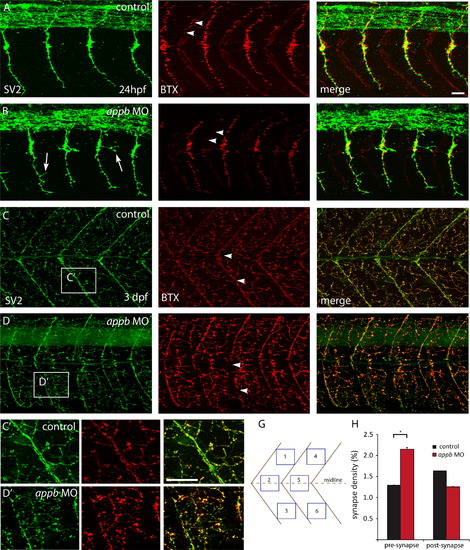
Increased pre-synapses and decreased post-synapses. Neuromuscular synapses are labeled with antibodies against SV2 (green, pre-synaptic vesicles) and αBTX (red, post-synaptic AChRs). Images are oriented so that left is rostral and top is dorsal. At 24 hpf, PMN of wildtype embryos (n=8) extends straight axons projecting dorsally and ventrally has established robust and overlapping NMJs (A). In appb morphants (n=8,) the ventral projections are branched (arrows) and have less post-synapses at the horizontal myoseptum (arrowheads) compared to control (B). At 3 dpf there is an elaborative network of pre- and post-synapses. Control larvae have well defined NMJs at the horizontal myoseptum (arrowheads) and evenly distributed NMJs in the somites (C). The appb morphants (D) have interrupted synapse formation at the horizontal myoseptum (arrowheads) and at the midline. (C′, D′) Higher magnification of C and D. (H) Measurement of the density of pre- and post-synapses were made by counting 6 areas (G) distributed dorsally, ventrally or at the midline of two somites of each fish. Scale bars: A, C 20 μm.
|
