FIGURE
Fig. 4
Fig. 4
|
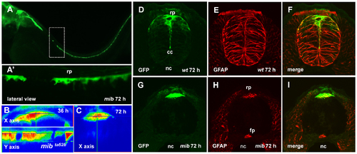
The roof plate formation in the mib mutant. (A), Confocal images, lateral view of the spinal cord of mib mutant, 72 hpf. Dashed rectangular shows the magnified in (A2) region of the spinal cord with the absence of GFP-positive RP cells. (B, C), the orthogonal optical sections of confocal images of the spinal cord of mib mutant illustrate lack of the roof plate extension between 36 and 72 hpf. Immunofluorescent staining of the transverse sections of spinal cord using anti-GFP (green) and anti-GFAP (red) antibodies; wild-type embryo (D–F) and mib mutant (G–I). |
Expression Data
| Gene: | |
|---|---|
| Antibody: | |
| Fish: | |
| Anatomical Terms: | |
| Stage: | Protruding-mouth |
Expression Detail
Antibody Labeling
Phenotype Data
| Fish: | |
|---|---|
| Observed In: | |
| Stage: | Protruding-mouth |
Phenotype Detail
Acknowledgments
This image is the copyrighted work of the attributed author or publisher, and
ZFIN has permission only to display this image to its users.
Additional permissions should be obtained from the applicable author or publisher of the image.
Full text @ PLoS One

