Fig. 8
- ID
- ZDB-FIG-121205-56
- Publication
- Biacchesi et al., 2012 - Both STING and MAVS Fish Orthologs Contribute to the Induction of Interferon Mediated by RIG-I
- Other Figures
- All Figure Page
- Back to All Figure Page
|
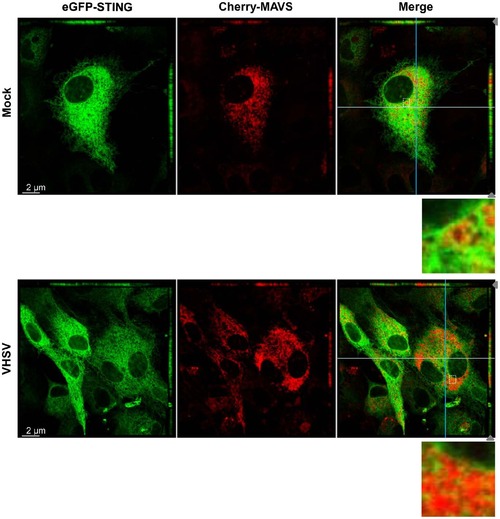
STING and MAVS are in close vicinity in mitochondrial-ER contact regions. EPC cells were cotransfected with 1 µg of peGFP-STING and 1 µg of pCherry-MAVS. At 48 h posttransfection, EPC were infected with VHSV at an MOI of 1 and incubated at 15°C for 24 h before imaging by confocal microscopy. For both panels the main images show a section of the cell monolayer in the xy plane at the z position indicated by the grey arrow head in the xz plane (small top panel) and the yz plane (small right side panel). Orthogonal projections of confocal sections shown in the top and right side panels are in the cutting plane indicated by the white and the blue lines corresponding to the xz and yz planes, respectively. Zoomed insets of boxed areas in merged images are also presented. |

