FIGURE
Fig. 7
- ID
- ZDB-FIG-121205-55
- Publication
- Biacchesi et al., 2012 - Both STING and MAVS Fish Orthologs Contribute to the Induction of Interferon Mediated by RIG-I
- Other Figures
- All Figure Page
- Back to All Figure Page
Fig. 7
|
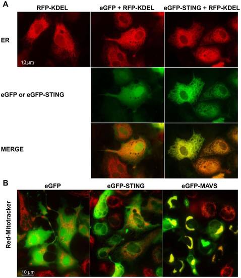
Localization of zebrafish STING to endoplasmic reticulum. peGFP-STING or peGFP-MAVS and peGFP, as negative controls, were transfected together with a plasmid encoding the Red Fluorescent protein fused to a reticulum endoplasmic location signal (RFP-KDEL) into EPC cells (A). The mitochondria were in vivo stained with a red MitoTracker (B). The cells were imaged by microscopy 24 h post-transfection. The yellow staining in the overlay image indicates colocalization of STING and RFP-KDEL (A) or MAVS and MitoTracker (B). |
Expression Data
Expression Detail
Antibody Labeling
Phenotype Data
Phenotype Detail
Acknowledgments
This image is the copyrighted work of the attributed author or publisher, and
ZFIN has permission only to display this image to its users.
Additional permissions should be obtained from the applicable author or publisher of the image.
Full text @ PLoS One

