
Fig. 4
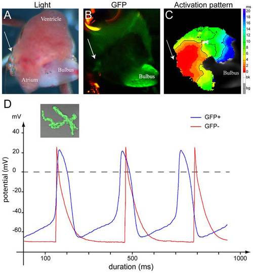
Isl1 cells have pacemaker activity. (A–C) Optical mapping on an explanted, contracting adult zebrafish tg(isl1BAC:GalFF; UAS:GFP) heart. Arrow indicates the sinus venosus in all panels. (A) Explanted adult zebrafish heart. (B) GFP-fluorescent cells reporting Isl1 expression are situated at the sinus venosus. (C) The activation pattern measured by di-4-ANEPPS fluorescence shows that the GFP+ myocytes are situated in the area of earliest activation. (D) Typical action potentials of freshly isolated GFP+ and GFP- myocytes. The GFP- cell was stimulated at 3 Hz. The inset displays a representative example of a GFP+ myocyte. |
| Gene: | |
|---|---|
| Fish: | |
| Anatomical Term: | |
| Stage: | Adult |