Fig. S4
- ID
- ZDB-FIG-100903-52
- Publication
- Wang et al., 2010 - Moesin1 and Ve-cadherin are required in endothelial cells during in vivo tubulogenesis
- Other Figures
- All Figure Page
- Back to All Figure Page
|
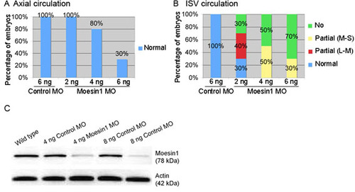
Moesin1 knockdown embryos display dose-dependent MO-induced defects in vascular perfusion. Ten embryos per group injected with TMRD at 54 hpf were scored for vascular perfusion for each experiment. Results are shown for one out of three similar experiments. (A) The percentage of embryos showing normal circulation in the dorsal aorta and posterior cardinal vein. A dose-dependent reduction of axial circulation is observed after injection of the moesin1 MO. (B) The percentage of embryos displaying either normal, partial (L-M, low to medium: 5-50% ISVs have no blood circulation), partial (M-S, medium to strong: 50-90% ISVs have no blood circulation), or no circulation (>90% ISVs have no blood circulation) in the ISVs after injection of either a control or moesin1 MO. The moesin1 MO was injected at 2, 4 and 6 ng and displays dose dependence for producing defects in circulation. (C) Western blot analysis of Moesin1 using an anti-Moesin antibody (BD Biosciences) following injection of either a control or moesin1 MO. Three embryos at 30 hpf were loaded per lane. The blot was reprobed with a mouse anti-β-Actin antibody (Sigma) as a loading control. Moesin1 MO dramatically reduces the level of Moesin1 protein. |

