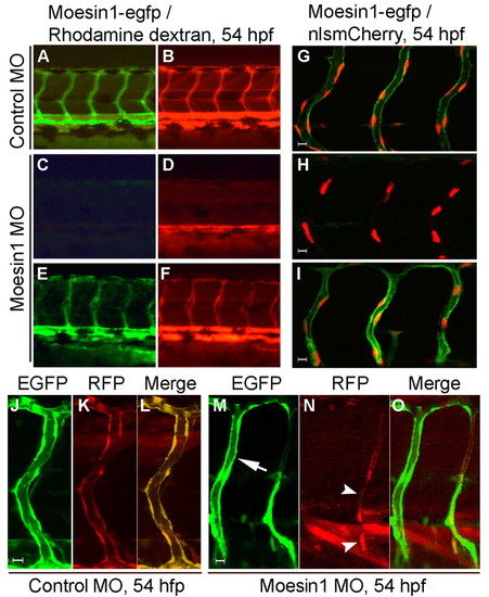
Moesin1 is required in the endothelial cells for tubulogenesis. (A-F) The Tg(flk1:moesin1-egfp) line partially rescues the Moesin1 knockdown phenotype. (A,B) A Tg(flk1:moesin1-egfp) zebrafish embryo injected with 4 ng control MO. All the ISVs were perfused with tetramethylrhodamine-dextran (TMRD, red). (C,D) Wild-type sibling embryo injected with 4 ng moesin1 MO. Most ISVs are not perfused with TMRD, although circulation of TMRD is observed in the axial vessels. (E,F) A Tg(flk1:moesin1-egfp) embryo injected with 4 ng moesin1 MO. Most ISVs are perfused with TMRD. (G-I) Same experiment as in A-F in Tg(flk1:moesin1-egfp)/Tg(flk1:nlsmCherry) embryos shows that the endothelial cells are present in the ISVs despite a lack of circulation at 54 hpf. (J-O) Transplantation of endothelial cells was used to determine autonomous verses non-autonomous effects. (J-L) Normal endothelial tubulogenesis occurs when the donor embryo was injected with control MO. (M-O) Transplanted endothelial cells from Moesin1 knockdown embryos fail to undergo normal tubulogenesis (arrowheads), whereas the ISV in the recipient embryo (arrow) completes lumen formation.
|

