
Fig. S4
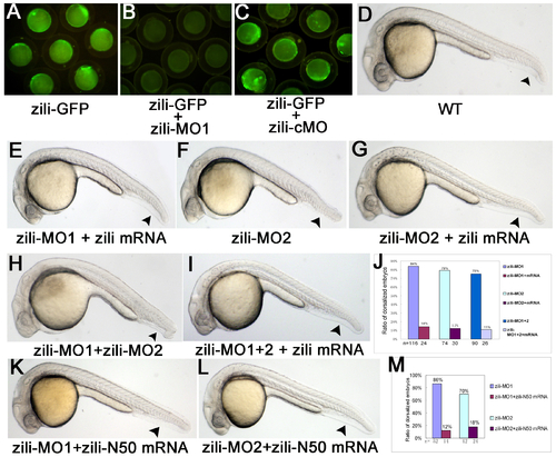
Specification and effectiveness of zili morpholinos. (A-C) Live embryos at the 70%-epiboly stage. (D-I, K-L) Lateral view of live embryos at 24 hpf, anterior to the left. Embryos injected with 100 pg pZili-GFP DNA produced green fluorescent fusion protein (A). Expression of the fusion protein was inhibited by coinjecting with 5 ng zili-MO1 (B), but not by coinjecting with 5 ng control morpholino (zili-cMO) (C). (D)Wild-type. (E) Embryos coinjected with 5ng zili-MO1 and 30 pg zili mRNA. Injection with 40 ng zili-MO2 (F) or zili-MO1+2 (consisting of 3 ng zili-MO1 and 20 ng zili-MO2) (H) caused the dorsalized phenotypes same to zili-MO1 and 30 pg zili mRNA also can relieve the morphological changes resulted from zili-MO2 or zili-MO1+2 (G and I). (J and M) The ratios of dorsalized embryos injected with zili morpholinos. The number of calculated embryos is indicated below each bar. Arrowhead in (D-I, K-L) shows the tail fin and tail. |