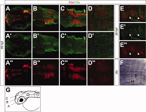
Dla is located in a regionally restricted pattern in the hindbrain. All images show dorsal views of 48 hours postfertilization (hpf; A-D″,F) and 28 hpf (E-E″) embryos. A-E″ are labeled with Elavl (red) and Dla (green) antibodies; F shows dla mRNA distribution. A′,B′,C′,D′,E′ show Dla staining, A″,B″,C″,D″,E″ show Elavl staining. A-A″: In the ventral part of the developing brain, Dla is detected in cell clusters near the midline. B-C″: Medial and dorsal Dla distribution is close to the midline and next to the rhombomere borders. D-D″: Dla labeling shows a regular pattern in the hindbrain, exclusive of Elavl staining. E-E″: Partial view of three rhombomeres at 28 hpf. Arrows mark rhombomere boundaries. Dla is located next to rhombomere borders whereas Elavl is expressed within the rhombomeres. F: dla mRNA expression resembles the Dla protein labeling shown in (E-E′). Black arrows mark dla-positive strings of cells in both anteroposterior axis and mediolateral axis. G: Cartoon showing the relative level of the images in A, B, and C. Scale bars = 53 μm in A-C″, 46 μm in D-D″,F, 25 μm in E-E″.
|
