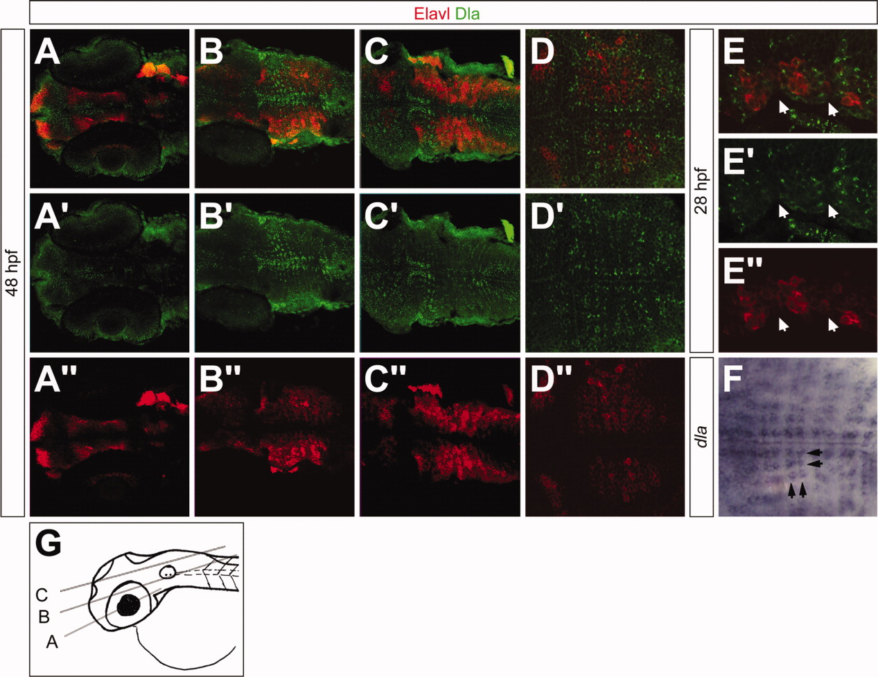
Fig. 4 Dla is located in a regionally restricted pattern in the hindbrain. All images show dorsal views of 48 hours postfertilization (hpf; A-D″,F) and 28 hpf (E-E″) embryos. A-E″ are labeled with Elavl (red) and Dla (green) antibodies; F shows dla mRNA distribution. A′,B′,C′,D′,E′ show Dla staining, A″,B″,C″,D″,E″ show Elavl staining. A-A″: In the ventral part of the developing brain, Dla is detected in cell clusters near the midline. B-C″: Medial and dorsal Dla distribution is close to the midline and next to the rhombomere borders. D-D″: Dla labeling shows a regular pattern in the hindbrain, exclusive of Elavl staining. E-E″: Partial view of three rhombomeres at 28 hpf. Arrows mark rhombomere boundaries. Dla is located next to rhombomere borders whereas Elavl is expressed within the rhombomeres. F: dla mRNA expression resembles the Dla protein labeling shown in (E-E′). Black arrows mark dla-positive strings of cells in both anteroposterior axis and mediolateral axis. G: Cartoon showing the relative level of the images in A, B, and C. Scale bars = 53 μm in A-C″, 46 μm in D-D″,F, 25 μm in E-E″.
Image
Figure Caption
Figure Data
Acknowledgments
This image is the copyrighted work of the attributed author or publisher, and
ZFIN has permission only to display this image to its users.
Additional permissions should be obtained from the applicable author or publisher of the image.
Full text @ Dev. Dyn.

