Fig. 2
- ID
- ZDB-FIG-091116-16
- Publication
- Suster et al., 2009 - A novel conserved evx1 enhancer links spinal interneuron morphology and cis-regulation from fish to mammals
- Other Figures
- All Figure Page
- Back to All Figure Page
|
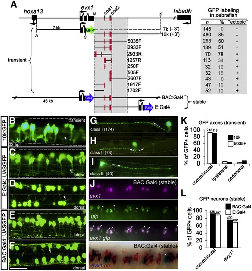
The pufferfish evx1 genomic region contains a downstream enhancer that labels evx1+ spinal commissural interneurons in zebrafish embryos. (A) Pufferfish evx1 locus and reporter constructs. Left, schematic of pufferfish evx1 reporter constructs. Black boxes represent exons. Translation start and orientation is indicated by arrows. For transient expression, farnesylated GFP and polyA (not shown) was fused in frame with exon 2 of evx1 (excluding homeodomain). 7 k contains the upstream promoter and lacks 3′ sequences (- 3′). 10 k contains a 10 kb downstream fragment (+ 3′) and others PCR fragments (size given in bp) downstream of GFP. Orientation is given as F (forward) or R (reverse). Restriction sites used were A (ApaI), N (NotI), S (SmaI) and X (XhoI). For stable transgenes, Gal4 was fused in frame with pufferfish evx1 using a ∼ 65 kb BAC and plasmid constructs (see Materials and methods). Right, observed frequencies of transient labeling in 28–32 hpf embryos. n, GFP+ embryos, and %, proportion with GFP+ interneurons. Frequent labeling of non-neuronal cells is noted under “ectopic”. (B–F) GFP-labeled interneurons (32 hpf) at rostral spinal cord levels with pufferfish evx1 constructs. B, C, E are lateral views and D, F are dorsal views. ∼ 2.5 segments are shown in each image. The limits of the spinal cord are indicated in B, C, E and the midline in D, F by dashed lines. Dorsal is up and rostral is to the left. Most neurons extend an axon (arrowhead in B, C) ventrally before crossing to the opposite side (out of focus). Scale bar: 35 μm. (G, H, I) Single-cell morphology of spinal interneurons labeled by 10k:GFP and 5035F:GFP constructs. (G) Class I (174 cells), commissural interneuron with large dorsal soma. Axon extends ventrally, crosses and then ascends dorsally 2–5 segments (arrow). (H) Class II (74 cells), commissural interneuron with medial soma whose axon extends ventrally before crossing and bifurcating (arrows). (I) Class III (40 cells), commissural interneuron with medial soma and a descending axon (arrow). (J) Dual ISH of evx1 (magenta) and gfp (green) mRNA in the spinal cord of stable transgenic BAC:Gal4; UAS:GFP embryos (fluorescent ISH, top panel and chromogenic ISH, bottom panel). Asterisks mark gfp+ cells that do not co-express evx1 and arrowheads mark cells labeled by evx1. In the bottom panel, a white arrowhead marks a cell that expresses only evx1. (K) Quantitation of axonal morphology of GFP+ spinal neurons labeled by 10k:GFP (10 k) and 5035F:GFP (5035F) in transient assays (shown in B). GFP+ cells were grouped as commissural, ipsilateral or peripheral based on the axonal projection. The total number of GFP+ cells is indicated above the bars. (L) Quantitation of axonal morphology and evx1 mRNA co-expression of spinal neurons labeled in BAC:Gal4; UAS:GFP and E:Gal4; UAS:GFP double transgenic embryos. Percentage (%) of GFP+ spinal neurons with commissural axons and gfp+ cells double labeled by evx1 mRNA are plotted. The total number of cells is indicated above the bars. |
| Genes: | |
|---|---|
| Fish: | |
| Anatomical Terms: | |
| Stage: | Prim-15 |
Reprinted from Developmental Biology, 325(2), Suster, M.L., Kania, A., Liao, M., Asakawa, K., Charron, F., Kawakami, K., and Drapeau, P., A novel conserved evx1 enhancer links spinal interneuron morphology and cis-regulation from fish to mammals, 422-433, Copyright (2009) with permission from Elsevier. Full text @ Dev. Biol.

