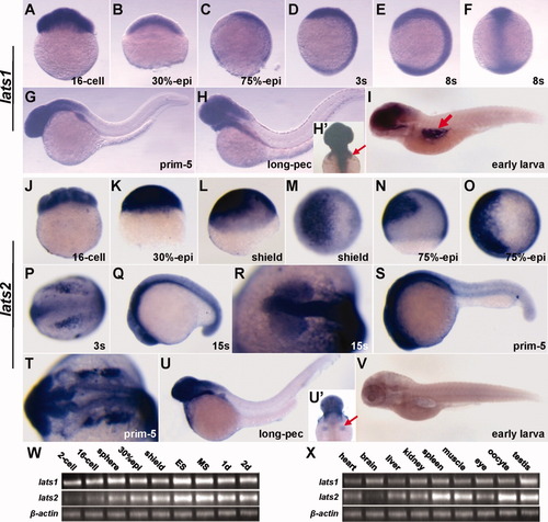
Expression patterns of lats1 and lats2 during development and in adult tissues of zebrafish. A-I: Expression analysis of lats1 by whole-mount in situ hybridization (WISH) in embryogenesis. A: lats1 is maternally expressed as shown by the strong staining in 16-cell stage embryo. B-F: lats1 is ubiquitously expressed during later development until mid-somitogenesis. G,H: From prim-5 to long-pec stage, lats1 is prominently expressed in the anterior structures such as the brain and pectoral fins (indicated by red arrow in panel H′). I: At early larva stage, as indicated by red arrow, lats1 is remarkably expressed in the gas bladder. J-V: Expression analysis of lats2 by WISH in embryogenesis. J,K: Similar to lats1, lats2 is likewise maternally and ubiquitously expressed before 30%-epiboly. L-T: From shield stage, lats2 begins to present territory-specific expression patterns, besides a low-level ubiquitous expression throughout these developmental stages. L-O: From shield (L,M) to mid-gastrula (N,O) stage, lats2 probe strongly labeled the non-neural ectoderm cells. P-T: At the three-somite stage (P), lats2 is mainly expressed in the neural crest cells and the primordial CNS (central nervous system) cells; from mid-somitogenesis (Q,R) to prim-5 (S,T) stage, lats2 is notably expressed in the eyes and all the parts of encephalon as well as the neural crest derivatives. U: At long-pec stage, lats2 is prominently expressed in the anterior structures such as the brain and pectoral fins (indicated by red arrow in panel U′). V: At the early larva stage, in contrast to lats1, there is no obvious expression of lats2 in the gas bladder. W: By Reverse transcription polymerase chain reaction (RT-PCR) analysis, lats1 is ubiquitously expressed from the two-cell to long-pec stage, and in comparison to lats1, lats2 shows weaker expression at 2-cell stage and stronger expression from early-somitogenesis. X: In different tissues of adult, lats1 is almost ubiquitously expressed and lats2 is more prominently expressed in the spleen, muscle and gonads. Embryos in A-E,J-L,N are lateral view with dorsal to the right; embryo in F is dorsal view with animal pole to the top; embryos in M,O are animal pole view with dorsal to the right; embryos in P,R,T are dorsal view with anterior to the left; embryos in G-I,Q,S,U,V are lateral view with anterior to the left; embryos in H′,U′ are dorsal view with anterior to the top.
|
