Fig. S5
- ID
- ZDB-FIG-090306-32
- Publication
- Löhr et al., 2009 - Zebrafish diencephalic A11-related dopaminergic neurons share a conserved transcriptional network with neuroendocrine cell lineages
- Other Figures
- All Figure Page
- Back to All Figure Page
|
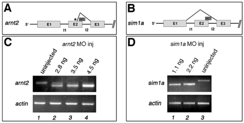
arnt2 and sim1 morpholinos effectively block correct splicing of pre-mRNA. (A) Schematic figure showing the first three exons and introns of the arnt2 gene and the splice morpholino targeting the exon2-intron2 boundary used in this study. A cryptic splice site in exon2 (depicted by an asterisk) causes deletion of 19 basepairs of the 3′ end of exon2, leading to a frameshift and a truncated Arnt2 protein. (B) Schematic figure showing the first three exons and introns of the sim1a gene and the splice morpholino targeting the exon2-intron2 boundary of sim1a. The morpholino leads to a complete loss of exon2 (83 basepairs). (C) RT-PCR from uninjected embryos (lane 1) and from embryos injected with 2.8 ng (lane 2), 3.5 ng (lane 3) or 4.5 ng (lane 4) arnt2 splice morpholino showing dose-dependent downregulation of wild-type splice pattern arnt2 transcript in injected embryos. The amount of actin transcript is unchanged. (D) RT-PCR from uninjected embryos (lane 1) and from embryos injected with 1.1 ng (lane 2) or 2.2 ng (lane 3) sim1a splice morpholino showing dose-dependent downregulation of wild-type splice pattern sim1a transcript in injected embryos. The amount of actin transcript is unchanged. |

