FIGURE

Fig. 8

ID
ZDB-FIG-090304-63
Publication
Feijoo et al., 2009 - Formation of posterior cranial placode derivatives requires the Iroquois transcription factor irx4a
Other Figures
All Figure Page
Back to All Figure Page
Fig. 8

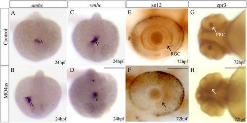
irx4a is involved in cardiac chamber specification and retinal development. Control and irx4a morphant embryos were processed for in situ hybridization (A–D) or immunohistochemistry (E–H). (A–D) Morphant embryos (B, D) show an increase in the size of the atrium and a decrease in the size of the ventricle compared to control embryos (A, C) as revealed the expression of the amhc (A, B) and vmhc (C, D) markers, respectively. (E, F) zn12 immunostaining labels retinal ganglion cells (RGC) in control embryos (E, arrow) but they are not detected in MO4as injected embryos (F). (G, H) zpr-3 immunostaining labels photoreceptor cells (PRC) in the 72 hpf control larva (G, arrow) but not in the morphant larva (H); note that photoreceptors have only differentiated in the ventral retina at this stage. A–D, dorsal views of whole embryos at 24 hpf, anterior is down; E, F, lateral views of the eye at 72 hpf, anterior is left; G, H, ventral views of the head. Scale bar in D: 40 μm for A–D, scale bar in F: 7 μm for E, F; scale bar in H: 15 μm for G, H.

Expression Data
Genes:
Antibodies:
Fish:
Knockdown Reagent:
Anatomical Terms:
Stage Range: Prim-5 to Protruding-mouth

Expression Detail
Antibody Labeling
Phenotype Data
Fish:
Knockdown Reagent:
Observed In:
Stage: Prim-5

Phenotype Detail
Acknowledgments
This image is the copyrighted work of the attributed author or publisher, and ZFIN has permission only to display this image to its users. Additional permissions should be obtained from the applicable author or publisher of the image.

Reprinted from Molecular and cellular neurosciences, 40(3), Feijoo, C.G., Saldias, M.P., De la Paz, J.F., Gómez-Skarmeta, J.L., and Allende, M.L., Formation of posterior cranial placode derivatives requires the Iroquois transcription factor irx4a, 328-337, Copyright (2009) with permission from Elsevier. Full text @ Mol. Cell Neurosci.