FIGURE

Fig. S3

ID
ZDB-FIG-080605-3
Publication
Sidi et al., 2008 - Chk1 Suppresses a Caspase-2 Apoptotic Response to DNA Damage that Bypasses p53, Bcl-2, and Caspase-3
Other Figures
All Figure Page
Back to All Figure Page
Fig. S3

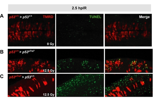
Validation of the cell transplantation assay
See Figure 3D for details on experimental procedure (cell transplantation and IR exposure).
(A) Dorsal view of a 5 μm thick confocal section of a p53+/+ spinal cord. TMR Dextran (red) marks cells from a p53+/+ donor embryo, which were transplanted at the blastula stage. TUNEL shown in green. The genetic chimera was not irradiated. Note that the transplanted p53+/+ cells do not stain TUNEL positive, showing that the transplantation technique does not induce apoptosis.
(B,C) 5 μm thick confocal sections of spinal cords in irradiated chimeras. TMR Dextran (red) marks the donor cells. TUNEL shown in green. (B), p53+/+ cells transplanted into a p53e7/e7 host. (C), p53e7/e7 cells transplanted into a p53+/+ host. Consistent with the fact that wild-type zebrafish embryos respond to IR through the cell autonomously-acting, mitochondrial apoptotic pathway (Berghmans et al., 2005; Kratz et al., 2006), p53+/+ cells transplanted into p53e7/e7 hosts stained TUNEL-positive after IR (86%, n=108) while neighboring p53e7/e7 cells remained largely TUNEL-negative (panel B). Furthermore, 86% (n=73) of p53e7/e7 cells transplanted into p53+/+ hosts remained refractory to IR-induced death, as indicated by their TUNEL negativity within an otherwise TUNEL-positive environment (panel C). Hence, TUNEL reactivity of transplanted cells after IR strictly depends on the p53 genotype of a cell, occurs irrespective of the cellular environment, and has very little, if any, influence on neighboring cells.

Expression Data

Expression Detail
Antibody Labeling
Phenotype Data

Phenotype Detail
Acknowledgments
This image is the copyrighted work of the attributed author or publisher, and ZFIN has permission only to display this image to its users. Additional permissions should be obtained from the applicable author or publisher of the image.

Reprinted from Cell, 133(5), Sidi, S., Sanda, T., Kennedy, R.D., Hagen, A.T., Jette, C.A., Hoffmans, R., Pascual, J., Imamura, S., Kishi, S., Amatruda, J.F., Kanki, J.P., Green, D.R., D'Andrea, A.A., and Look, A.T., Chk1 Suppresses a Caspase-2 Apoptotic Response to DNA Damage that Bypasses p53, Bcl-2, and Caspase-3, 864-877, Copyright (2008) with permission from Elsevier. Full text @ Cell