
Fig. S5
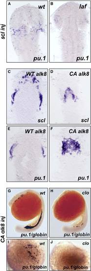
Genetic Epistatic Analysis of scl, cloche, and alk8 (A and B) lost-a-fin mutants injected with scl mRNA (50 ng/mL) did not display rescued expression of pu.1. Embryo in (A) is a wildtype sibling (n = 34) and in (B) a mutant sibling (n = 11). In the same experiment, scl mRNA injected from the same needle increased gata1 expression in posterior hematopoietic tissues (data not shown) as previously described [S18]. Embryo displayed as a flatmount, dorsal view of the anterior of the embryo. (C–F) Injection of CA alk8 mRNA does not increase scl expression. Embryos injected with WT alk8 mRNA (10 ng/mL) display normal expression of scl at 8 somites (C); embryos injected with CA alk8 mRNA (10 ng/mL) do not show an increase in scl expression at 8 somites (D) (n = 60/67 no increase [Class II], n = 7/67 vastly reduced anterior structures [Class III]). In the same experiment, CA alk8 mRNA-injected embryos displayed increased pu.1 expression (n = 62/105 increased) (F), but WT alk8 mRNA-injected embryos did not (E). Embryo displayed as a flatmount, dorsal view of the anterior of the embryo. (G–J) CA alk8 mRNA does not rescue pu.1 expression in cloche mutants. Wild-type siblings injected with CA alk8 mRNA, evidenced by mild ventralization, display normal expression of embryonic globin and pu.1 ([G], [I] [dorsal view], n = 46), whereas cloche mutants injected with CA alk8 mRNA, which were mildly ventralized, displayed vastly reduced globin expression and no expression of pu.1 (n = 24). Embryos in (G) and (H) are viewed laterally whereas (I) and (J) are views of the anterior. |