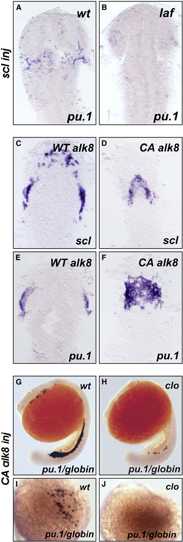
Fig. S5 Genetic Epistatic Analysis of scl, cloche, and alk8 (A and B) lost-a-fin mutants injected with scl mRNA (50 ng/mL) did not display rescued expression of pu.1. Embryo in (A) is a wildtype sibling (n = 34) and in (B) a mutant sibling (n = 11). In the same experiment, scl mRNA injected from the same needle increased gata1 expression in posterior hematopoietic tissues (data not shown) as previously described [S18]. Embryo displayed as a flatmount, dorsal view of the anterior of the embryo. (C–F) Injection of CA alk8 mRNA does not increase scl expression. Embryos injected with WT alk8 mRNA (10 ng/mL) display normal expression of scl at 8 somites (C); embryos injected with CA alk8 mRNA (10 ng/mL) do not show an increase in scl expression at 8 somites (D) (n = 60/67 no increase [Class II], n = 7/67 vastly reduced anterior structures [Class III]). In the same experiment, CA alk8 mRNA-injected embryos displayed increased pu.1 expression (n = 62/105 increased) (F), but WT alk8 mRNA-injected embryos did not (E). Embryo displayed as a flatmount, dorsal view of the anterior of the embryo. (G–J) CA alk8 mRNA does not rescue pu.1 expression in cloche mutants. Wild-type siblings injected with CA alk8 mRNA, evidenced by mild ventralization, display normal expression of embryonic globin and pu.1 ([G], [I] [dorsal view], n = 46), whereas cloche mutants injected with CA alk8 mRNA, which were mildly ventralized, displayed vastly reduced globin expression and no expression of pu.1 (n = 24). Embryos in (G) and (H) are viewed laterally whereas (I) and (J) are views of the anterior.
Image
Figure Caption
Acknowledgments
This image is the copyrighted work of the attributed author or publisher, and
ZFIN has permission only to display this image to its users.
Additional permissions should be obtained from the applicable author or publisher of the image.
Full text @ Curr. Biol.

