|
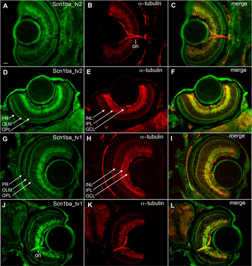
Retinal patterning of Scn1ba_tv1 and Scn1ba_tv2. A – F: anti-Scn1ba_tv2 (green), anti-acetylated α-tubulin (red). G – L: anti-Scn1ba_tv1 (green), anti-acetylated α-tubulin (red). Anti-Scn1ba_tv2 stains the layers of the retina, including the ganglion cell layer (GCL), inner plexiform layer (IPL), outer plexiform layer (OPL), outer limiting membrane (OLM), and photoreceptor cell layer (PR). Staining appears to be absent in the inner nuclear layer (INL) and in the optic nerve (on). Anti-Scn1ba_tv1 stains all the layers of the retina including the inner nuclear layer, where it shows robust staining. In contrast to anti-Scn1ba_tv2, anti-Scn1ba_tv1 labels optic nerve. Scale bar: 50 μm.
|