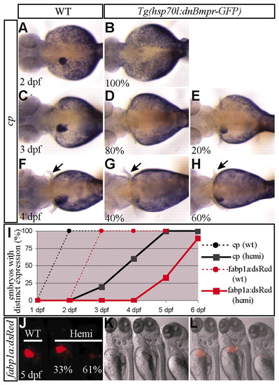
Response of foregut endodermal cells to a transient block in Bmp signaling. (A-H) Embryos obtained from outcrossing a hemizygous Tg(hsp70l:dnBmpr-GFP) zebrafish were heat shocked at 18 hpf for 25 minutes, harvested at 2 (A,B), 3 (C-E) or 4 (F-H) dpf and examined for cp expression. Distinct, hepatocyte expression of cp in the heat-shocked hemizygous embryos was not detected at 2 dpf (B), but was detected in 20% of the embryos at 3 dpf and 60% at 4 dpf (E,H). The percentage of hemizygous embryos exhibiting a similar expression level is indicated in the lower left corner (n=9-11). Arrows point to the pectoral fins. (I) The percentage of embryos exhibiting distinct, hepatocyte cp expression in A-H together with that of embryos at 5 and 6 dpf is shown in the graph (n=9-11). Black circles and black squares denote wild-type siblings and hemizygous embryos, respectively. Embryos obtained from crossing a hemizygous Tg(hsp70l:dnBmpr-GFP) fish with a homozygous Tg(fabp1a:dsRed) fish were treated in the same way as above, and examined for DsRed expression under a dissecting fluorescence microscope. Distinct, DsRed expression in the hemizygous embryos was not detected by 4 dpf, but was detected in 33% of the embryos at 5 dpf (J-L). Red circles and red squares denote wild-type siblings and hemizygous embryos, respectively (n=20). Fluorescence (J), brightfield (K) and a merged (L) image of the embryos at 5 dpf are shown. Embryos transiently lacking Bmp signaling eventually initiated cp and fabp1a- DsRed expression, although with a delay. A-H, dorsal views, anterior left; J-L, ventrolateral views, anterior up.
|
