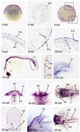
perp is expressed in specific cells of the zebrafish embryo. In situ hybridization with perp antisense riboprobe at different stages of development. (a,b) Shield stage (early gastrula; 6 hpf). (a) Whole-mount embryo, (b) Sagital section. (c-e) 80% epiboly (midgastrula; 8 hpf). (c) Whole mount embryo. (d,e) Longitudinal sections, anterior up, arrow in (e) indicates single deep cells at the dorsal margin. (f) Tailbud stage (end of gastrulation; 10 hpf), transverse section through dorsal midline, dorsal up. perp is expressed in the notochord anlage (nc). (g-k) 25-somite stage (24 hpf). (g) Lateral overview over entire embryo; positions of section (h), magnified region (i), and optical cross-section (j,k) are indicated. (h) Transverse section through the tail of embryo stained for perp mRNA (in blue) and p63 protein (in brown), close-up on the skin. (i) Magnified view on the tail tip. perp shows prominent expression in the notochord. (j) Optical cross-section at level of eyes. (j) Optical cross-section at the level of the otic vesicles. (k,l) 36 hpf. (l) Dorsal view on the head. (m) Transverse section through trunk. (n,o) 48 hpf. (n) Higher magnification of pectoral fin. (o) Dorsal view on trunk. Abbreviations: bae, branchial arch epithelium; bel, basal epidermal layer; EVL, enveloping layer; epi, epidermis, skin; gcl, ganglion cell layer of retina; gt, gut; hed, hepatic duct; le, lense; li, liver; nc, notochord; oel, outer epidermal layer; olf, olfactory epithelium of nasal pits; ov, otic vesicle; pan, pancreas; pcp, prechordal plate; pfc, pectoral fin cartilage; pn, pronephric duct; vdi, ventral diencephalon; vhb, ventral hindbrain
|

