FIGURE SUMMARY
Title

Knock-out of vasotocin reduces reproductive success in female zebrafish, Danio rerio

Authors
Ramachandran, D., Sharma, K., Saxena, V., Nipu, N., Rajapaksha, D.C., Mennigen, J.A.
Source
Full text @ Front Endocrinol (Lausanne)

Generation and validation of avp -/- zebrafish: (A) Microinjection and breeding scheme. (B) CRISPR/Cas9 construct and targeted locus. (C) DNA sequencing of avp locus in avp-/- fish (D) Agarose gel electrophoresis analyzing avp -/- transcript specific amplicons in brain and ovaries obtained by RT-PCR. Please refer to the text for detailed explanations.

Schematic representation of experimental design: (A) Breeding assays. (B) Investigation of mechanistic basis of the female reproductive phenotype using ovarian histology, quantification of ovarian hormone concentrations, and ovarian gene expression assays. (C) Single (1x) and repeated (3x over a period of 9d prior to breeding assay) vasotocin injection in avp-/- females prior to breeding to assess possible rescue of the reproductive phenotype. Please refer to the text for detailed explanations.

Assessment of reproductive success in WT (white bars and data points) and avp-/- (purple bars and data points) zebrafish. (A) Percent successful breeding trials in WT and avp-/- breeding pairs analyzed by Fisher’s exact test. (B) (Median) number of fertilized eggs in WT and avp-/- in all breeding trials analyzed by Mann-Whitney U test. Asterisk indicates a significant difference at P<0.01. (C) (Mean) clutch size expressed as the number of fertilized eggs in successful breeding trials analyzed by t-test. Asterisk indicates a significant difference at P<0.001. (D) Full-factorial crossbreeding experiments to delineate sex-specific contributions of the avp genoptype to reproductive success. Four crosses were bred: WT females x WT males; WT females x avp -/- males; WT males x avp -/- females; avp-/- males x avp-/- females. (D) (Median) number of fertilized eggs of each breeding trial across breeding groups analyzed by two-way ANOVA on ranks followed by Scheirer-Ray-Hare extension. (E) (Mean) Clutch size expressed as the number of fertilized eggs in successful breeding trials. Data were analyzed by two-way ANOVA (F).

Quantification of courtship behaviors in WT and avp -/- fish breeding pairs. The first 10 min of interaction were videotaped and analyzed by a researcher blind to the genotype of the breeding pairs. (A) (Median) number of nudges. (B) (Mean) number of chasing events. (C) (Mean) duration of chasing events, (D) (Median) number of circling events. (E) (Median) number of quivering events are shown. Data were analyzed by 2-way ANOVA where normally distributed or by two way-ANOVA on ranks followed by Scheirer-Ray-Hare extension for non-normally distributed data. The asterisk represents a significant effect of the maternal genotype, in which breeding pairs with avp -/- females exhibit significantly less quivering behaviour compared to pairings with female WT.

(A) (Mean) gonadosomatic index of WT (n=25) and avp -/- (n=26) females. (B, C) (Median) total oocyte number, and oocytes staged from ovarian histology sections of WT (n=4, 120 total sections) and avp-/- (n=5, 150 total sections). (D) Representative ovarian histology sections form WT and avp-/- females maximum diameter measurements to determine oocyte stage distribution as (mean) percentage (± S.E.M.) of overall oocytes according to the classification system described by Li and Ge (2020). Data were analyzed by t-test in case of normal distribution and Mann-Whitney U test in case of non-normal distribution. Asterisks indicate significant differences (P<0.001 in (B); P<0.05 in (C, D). (E) Ovarian histology sections from gravid WT (top panel) and avp-/- fish (bottom panel) imaged using a stereomicroscope (left images) and light microscope respectively (right images).

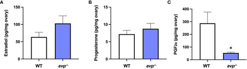
Mean ovarian sex steroid concentrations (± S.E.M.) of (A) E2, (B) P4, and (C) PGF in WT (n=18) and avp-/- (n=18) fish. Data were analyzed by t-test and significant differences (P<0.05) are indicated by asterisk.

Targeted gene expression analysis of selected transcripts with characterized roles in different stages of oocyte development in WT (n=7) and avp -/- (n=7) ovaries. Data are presented as mean values (± S.E.M.) normalized relative to WT fish transcript abundance to visualize fold-change Data were analyzed by t-test and significant differences (P<0.05) are indicated by asterisk.

Rescue experiments assessing reproductive success in female avp-/- fish following single 1x acute (A, B) or 3x repeated i.p. injections (every 3 d) (C, D) of physiological saline or 1 μg/g vasotocin prior to breeding assays with WT males. For the 1x acute injection experiment, n=15 avp-/- females were injected with physiological saline, and n=15 with 1 μg/g vasotocin prior to breeding assays with WT males. For the 3x repeated injection experiment, n=9 avp -/- females were injected with physiological saline, and n=8 avp -/- females were injected with 1 μg/g vasotocin prior to breeding assays with WT males. (A, C) Percentage of successful breeding pairs. (B, D) Median number of fertilized eggs of avp-/- fish used in breeding assays following i.p. injection of physiological saline or 1 μg/g 1 μg/g vasotocin. Data were analyzed by Fisher’s exact test and Mann-Whitney U test, respectively, and significant differences at P<0.05 are indicated by asterisk.

(A) Larval cortisol concentration at 5 dpf and (B) baseline cortisol concentration in sexually mature adult blood samples. Larval cortisol was determined in WT and avp-/- 5 dpf larvae pools (n=4 per genotype, 20 larvae per pool) according to published protocols (50). Adult whole blood cortisol was analyzed in adults of mixed sex and analyzed under baseline conditions for both WT (n=10) and avp-/- mutants (n=10) according to published protocols (51) with the modification that cortisol was extracted from whole blood. Data are presented as means ± S.E.M. and were analyzed using a t-test. A threshold of P<0.05 was used to discern a significant difference, which is indicated by asterisk.

Acknowledgments
This image is the copyrighted work of the attributed author or publisher, and ZFIN has permission only to display this image to its users. Additional permissions should be obtained from the applicable author or publisher of the image. Full text @ Front Endocrinol (Lausanne)