FIGURE SUMMARY
Title

The blood flow-klf6a-tagln2 axis drives vessel pruning in zebrafish by regulating endothelial cell rearrangement and actin cytoskeleton dynamics

Authors
Wen, L., Zhang, T., Wang, J., Jin, X., Rouf, M.A., Luo, D., Zhu, Y., Lei, D., Gregersen, H., Wang, Y., Wang, G.
Source
Full text @ PLoS Genet.

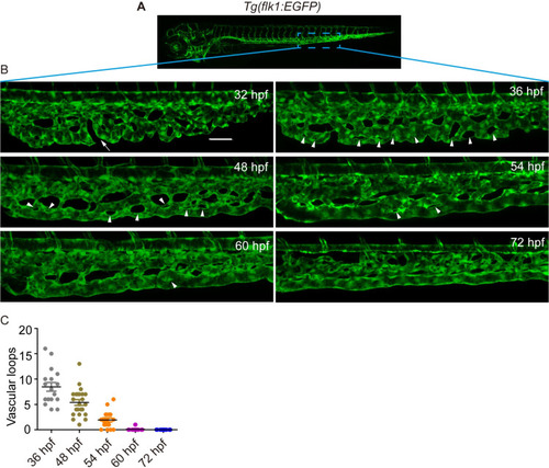
The zebrafish caudal vein formation involves vascular remodeling.

(A) A sketch map of the region. (B) Confocal images of the caudal vasculature of zebrafish embryos. The arrow points to ventral CVP capillaries without connection. Arrowheads point to vascular loops. Scale bar: 50 μm. (C) Quantification of the number of vascular loops in the CV: 36 hpf, n = 17 embryos; 48 hpf, n = 23 embryos; 54 hpf, n = 19 embryos; 60 hpf, n = 22 embryos; 72 hpf, n = 13 embryos.

EXPRESSION / LABELING:
Gene:
Fish:
Anatomical Term:
Stage Range: Prim-15 to Protruding-mouth

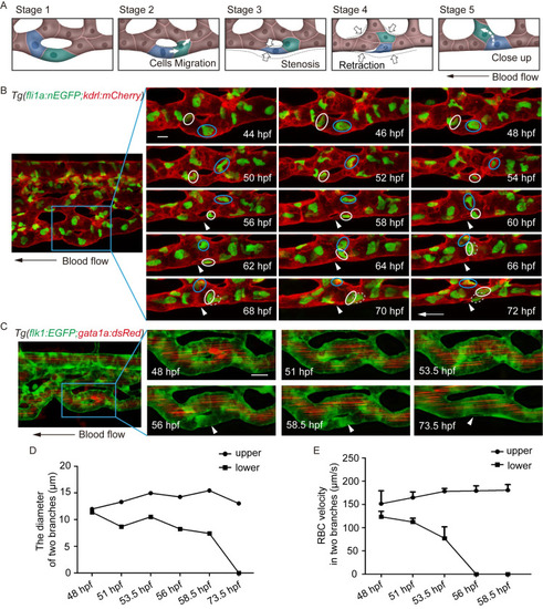
(A) Sketch map of EC rearrangement during vessel pruning. (B) Time-lapse live imaging of Tg(fli1a:nEGFP;kdrl:mCherry) embryos shows EC nucleus migration in CV pruning. Arrowheads indicate the pruned vessel. The arrows indicate the direction of the blood flow. Colored circles indicate ECs nuclei. 24 time-lapse live imaging were taken. Scale bar: 10 μm. (C) Time-lapse live imaging of Tg(flk1:EGFP;gata1a:dsRed) embryos shows vessel stenosis and the change of blood flow in CV pruning. Arrowheads indicate the lack of blood flow in the regressing vessel. The arrow indicates the direction of the blood flow. 10 time-lapse live imaging were taken. Scale bar: 10 μm. (D) The diameters of two branches at the indicated stages. (E) The velocity of RBCs in two branches at the indicated stages (48 hpf: S1 Video, 51 hpf: S2 Video, 53.5 hpf: S3 Video, 56 hpf: S4 Video, 58.5 hpf: S5 Video). The number of RBCs is calculated for blood flow at each stage: upper branch, 48.5 hpf: n = 5, 51 hpf: n = 10, 53.5 hpf: n = 8, 56 hpf: n = 9, 58.5 hpf: n = 10; lower branch, 48.5 hpf: n = 10, 51 hpf: n = 9, 53.5 hpf: n = 2.

Transcription factor <italic toggle='yes'>klf6a</italic> regulates CV pruning in zebrafish.

(A) Generation of KI (klf6a -HA-P2A-gal4) fish. (B) Expression pattern of KI(klf6a-HA-P2A-gal4) fish. Arrowheads indicate the location of klf6a at the vasculature. (C) Role of klf6a in CV pruning in zebrafish embryos. Boxes show enlarged images of the CV. The arrowhead indicates the unpruned vessel. Scale bar: 50 μm. (D) Quantification of vascular loops in the sibling and klf6a-/-: sibling, n = 23 embryos; klf6a-/-, n = 13 embryos. P < 0.001. Student’s unpaired two-tailed t test.

<italic toggle='yes'>Klf6a</italic> regulates EC nucleus migration in CV pruning.

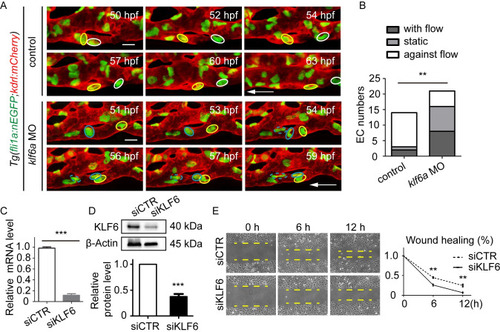
(A) Time-lapse live imaging of Tg(fli1a:nEGFP;kdrl:mCherry) zebrafish embryos shows EC nucleus migration in CV pruning in control and klf6a morphant. The arrow shows the direction of the blood flow. Colored circles indicate EC nuclei. Scale bar: 10 μm. (B) The direction of EC migration within the regressing vessel. The direction is classified into three types: with the flow, static, and against the flow. A total of 10 vascular loops with 14 EC nuclei and 14 vascular loops with 21 EC nuclei were calculated in the control group and klf6a morphant group, respectively. P = 0.0058. Unpaired two-tailed chi-square test. (C) and (D) Efficiency of siKLF6 in human umbilical vein endothelial cells (HUVECs) at the mRNA (P = 0.00087) and protein (P = 0.0003) levels. (E) Wound healing of siCTR-transfected and siKLF6-transfected HUVECs: 6 h, P = 0.0013; 12 h, P = 0.0020. (C) to (E) Student’s unpaired two-tailed t test. *P < 0.05, **P < 0.01, ***P < 0.001.

Knockdown of <italic toggle='yes'>klf6a</italic> impairs junction remodeling and rearrangement of actin cytoskeleton in CV pruning.

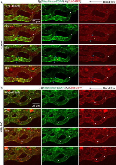
(A) and (B) Time-lapse live imaging of junction remodeling and actin cytoskeleton dynamics in CV pruning in Tg(Fliep:Lifeact-EGFP);KI(cdh5-mRFP) embryos. (A) Control embryo shows a retraction of junction (white arrowhead) and a shrinkage of its junctional ring (white and blue arrowheads) as multicellular tube became stenotic. F-actin forms at cdh5-positive junction (white, blue and yellow arrowheads), and undergoes a similar rearrangement as cdh5-positve junctions. 7 vascular loops are taken time-lapse live imaging. Scale bar: 25 μm. (B) Although klf6a morphant show F-actin co-localization with junction, however, neither junction nor F-actin does not go through remodeling (arrowhead). 5 vascular loops are taken time-lapse live imaging. Scale bar: 25 μm.

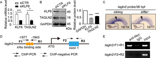
<italic toggle='yes'>Tagln2</italic> is a direct downstream target of <italic toggle='yes'>klf6a</italic>.

(A) and (B) SiKLF6 in HUVECs efficiently decrease TAGLN2 expression at the mRNA (KLF6: P = 0.0009, TAGLN2: P = 0.0352) and protein (KLF6: P < 0.0001, TAGLN2: P < 0.0001) levels. Student’s unpaired two-tailed t test. *P < 0.05, ***P < 0.001. (C) WISH of the tagln2 gene of sibling and klf6a-/- zebrafish at 36 hpf. Arrowheads indicate the CVP region expressing tagln2. (D) Structure of the tagln2 promoter. The primer tagln2-F1+R1 contains the conserved Klf6a binding side-CACCC, whereas tagln2-F2+R2 is a negative control located in the tagln2 exon 1. (E) ChIP-PCR of the tagln2 promoter shows that Klf6a can bind directly to the tagln2 promoter.

EXPRESSION / LABELING:
Gene:
Fish:
Anatomical Term:
Stage: Prim-25
PHENOTYPE:
Fish:
Observed In:
Stage: Prim-25

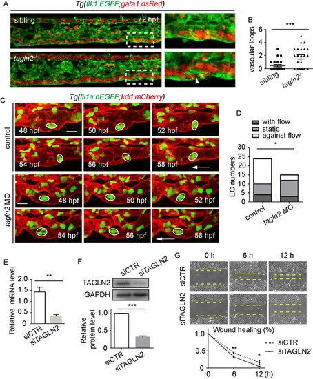
<italic toggle='yes'>Tagln2</italic> regulates CV pruning by promoting EC nucleus migration.

(A) Role of tagln2 in CV pruning in zebrafish embryos. Boxes show enlarged images of the CV. The arrowhead indicates unpruned vessel. Scale bar: 50 μm. (B) Quantification of vascular loops in sibling and tagln2-/- embryos: sibling, n = 25 embryos; tagln2-/-, n = 23 embryos. P = 0.0004. Student’s unpaired two-tailed t test. (C) Time-lapse live imaging of Tg(fli1a:EGFP;kdrl:mCherry) embryos shows EC nucleus migration in CV pruning in control or tagln2 morphant. Arrows indicate the direction of the blood flow. Colored circles indicate the EC nuclei. Scale bar: 10 μm. (D) Direction of EC nucleus migration in the regressing vessel. The direction is classified into three types: with the flow, static, and against the flow. A total of 12 vascular loops with 24 EC nuclei and 8 vascular loops with 15 EC nuclei were calculated in the control group and tagln2 morphant, respectively. Unpaired two-tailed chi-square test. P = 0.0471. (E) and (F) Efficiency of siTAGLN2 in HUVECs at the mRNA (P = 0.0094) and protein (P < 0.0001) levels. (G) Wound healing of siCTR-transfected and siTAGLN2-transfected HUVECs. Statistics for wound healing after siCTR and siTAGLN2 transfection: 6 h, P = 0.0037; 12 h, P = 0.0252. (E)-(G) Student’s unpaired two-tailed t test. *P < 0.05, **P < 0.01, ***P < 0.001.

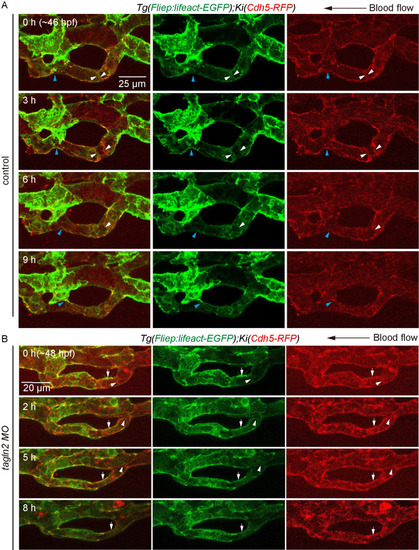
Knockdown of <italic toggle='yes'>tagln2</italic> impairs junction remodeling and rearrangement of F-actin cytoskeleton in CV pruning.

(A) and (B) Time-lapse live imaging of junction remodeling and actin cytoskeleton dynamics in CV prunig in Tg(Fliep:Lifeact-EGFP);KI(cdh5-mRFP) embryos. (A) Control embryo shows a detachment of junction (blue arrowheads) and an opposite direction of junction movement (white arrowheads) as multicellular tube became stenosis. F-actin forms at cdh5-positive junction (white and blue arrowheads), and undergoes a similar rearrangement as cdh5-positve junctions. Seven vascular loops are taken time-lapse live imaging at the stage of multicellular tube. Scale bar: 25 μm. (B) In tagln2 morphant, cdh5-positive junction moves to the right and connected with the adjacent one (white arrowheads), and F-actin undergoes similar rearrangement within it (white arrowheads). However, depolymerization of F-actin is also observed at junctions (white arrowheads). Six vascular loops are taken time-lapse live imaging. Scale bar: 20 μm.

Acknowledgments
This image is the copyrighted work of the attributed author or publisher, and ZFIN has permission only to display this image to its users. Additional permissions should be obtained from the applicable author or publisher of the image. Full text @ PLoS Genet.