FIGURE SUMMARY
Title

Loss of Pde6a Induces Rod Outer Segment Shrinkage and Visual Alterations in pde6aQ70X Mutant Zebrafish, a Relevant Model of Retinal Dystrophy

Authors
Crouzier, L., Diez, C., Richard, E.M., Cubedo, N., Barbereau, C., Rossel, M., Delaunay, T., Maurice, T., Delprat, B.
Source
Full text @ Front Cell Dev Biol

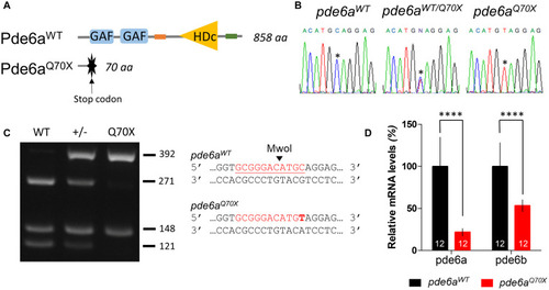
Mutation in pde6a zebrafish causes a premature stop codon. (A) Structure of zebrafish Pde6a protein with specific GAF (cGMP-specific phosphodiesterases, adenylyl cyclases and FhlA) and HDc [histidine (H) and/or aspartate (D) amino acid residues] domains. (B) Sanger sequencing identified the nucleotide mutation in exon 1 of pde6a sequence. Asterisk (*) denotes the substituted nucleotide in the knock-out pde6a sequencing trace. (C) A targeted fragment was amplified by PCR from the genomic DNA of adult zebrafish tail followed by an enzymatic digestion with Mwo1. The three rows present digested products for pde6aQ70X, pde6aWT, and pde6aWT/Q70X fish. Mwo1 restriction site is shown on the side of the agarose gel image. The mutation is highlighted in bold. (D) Expression level of pde6a and pde6b mRNA revealed by qPCR in pde6aQ70X and pde6aWT zebrafish larvae. Expression of pde6a and pde6b were normalized using the zef1α reference gene. Error bars represent SD calculated from three replicas. ****p < 0.0001.

EXPRESSION / LABELING:
Genes:
Fish:
Anatomical Term:
Stage: Day 5
PHENOTYPE:
Fish:
Observed In:
Stage: Day 5

Morphology of the pde6aQ70X larvae at 5-dpf. (A) Schematic representation of the different measurements of the larva (modified from Lizzy Griffiths). (B)pde6aQ70X larvae showed presence of swim bladders and absence of any morphological abnormality as compared to pde6aWT controls. (C) Measurement of body length, from the mouth to the end of the tail fin and (D) the diameter of the eye. The area of the ear (E), anterior (F), and posterior (G) otolith were also analyzed. Scale = 1 mm. The number of animals is indicated within the columns. p > 0.05 for all comparisons.

Impact of pde6a mutation on the OKR test of larvae at 5-dpf. (A) OKR chromograph illustrating the saccades performed by pde6aWT larvae (in blue) and pde6aQ70X larvae (in red) for 1 min. (B) Quantification of the number of saccades performed in 2 min. The number of animals is indicated within the columns. The experiment was repeated 3 times.

Analysis of the distance traveled by the larvae during the light/dark cycle in the VMR test at 5 dpf. (A) Sequence protocol: the activity is measured for 70 min, with 30 min of training in the dark (OFF), then 2 cycles of light/dark (ON/OFF) of 10 min each. (B) Average distance traveled per min for each condition according to the light/dark protocol. Distance traveled by the larvae during: (C) the averaged OFF1 and OFF2 phases; (D) the training phase [blue dotted lines in (B), between 21 and 29 min]; and (E) the averaged ON1 and ON2 phases. Distance were expressed as % of controls. Error bars represent the SD from three replicas except for B where errors bars represent the SEM. The number of animals is indicated within the columns. ∗∗p < 0.01.

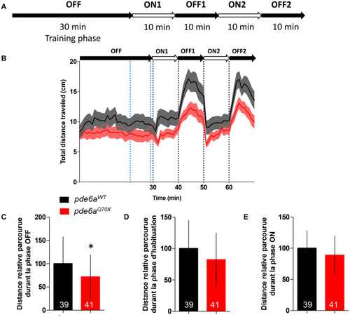
Analysis of the distance traveled by the larvae during the light/dark cycle in the VMR test at 21 dpf. (A) Sequence protocol: the activity is measured for 70 min, with 30 min of training in the dark (OFF), then 2 cycles of light/dark (ON/OFF) of 10 min each. (B) Average distance traveled per min for each condition according to the light/dark protocol. Distance traveled by the larvae during: (C) the averaged OFF1 and OFF2 phases; (D) the training phase [blue dotted lines in (B), between 21 and 29 min]; and (E) the averaged ON1 and ON2 phases. Distance were expressed as % of controls. Error bars represent the SD from three replicas except for B where errors bars represent the SEM. The number of animals is indicated within the columns. p < 0.05.

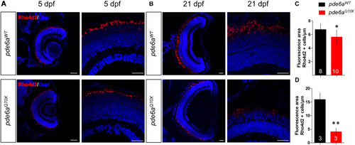
Analysis of rods in pde6aQ70X zebrafish during development. Representative confocal images of the retina of pde6aWT and pde6aQ70X zebrafish obtained after immunostaining of rods with a Rho4d2 antibody highlighting Rhodopsin (red) at 5 dpf (A) and 21 dpf (B). The nuclei were counterstained with DAPI (blue). Scale bar = 30 μm. (C,D) Quantification of the total area of rod outer segments normalized against the length of the retinal section, based on three independent measurements of larvae per group at 5 dpf (C) and 21 dpf (D). The number of animals is indicated within the columns. p < 0.05, ∗∗p < 0.01.

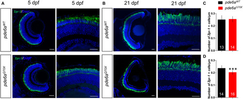
Analysis of cones in pde6aQ70X zebrafish during development. Representative confocal images of the retina of pde6aWT and pde6aQ70X zebrafish obtained after immunostaining of cones with a Zpr-1 antibody (green) at 5 dpf (A) and 21 dpf (B). Nuclei were highlighted with DAPI (blue). Scale bar = 30 μm. (C,D) Quantification of the number of cones normalized against the length of the retinal section, based on three independent measurements of larvae per group at 5 dpf (C) and 21 dpf (D). The number of animals is indicated within the columns. ∗∗∗p < 0.001.

Analysis of activated Caspase-3 positive cells in pde6aQ70X zebrafish during development. (A,B) Representative confocal images of the retina of pde6aWT and pde6aQ70X zebrafish obtained after immunostaining of the retinal section with activated Caspase-3 antibody (red) at 5 dpf (A) and 21 dpf (B). Nuclei were highlighted with DAPI (blue). Scale bar = 30 μm. (C,D) Quantification of the number of activated Caspase-3 positive cells in the entire retina based on three independent measurements of larvae per group at 5 dpf (C) and 21 dpf (D). The number of animals is indicated within the columns. ** p < 0.01, ***p < 0.001.

EXPRESSION / LABELING:
Antibody:
Fish:
Anatomical Term:
Stage Range: Day 5 to Days 21-29
Acknowledgments
This image is the copyrighted work of the attributed author or publisher, and ZFIN has permission only to display this image to its users. Additional permissions should be obtained from the applicable author or publisher of the image. Full text @ Front Cell Dev Biol