FIGURE SUMMARY
Title

Knockout of zebrafish desmin genes does not cause skeletal muscle degeneration but alters calcium flux

Authors
Kayman Kürekçi, G., Kural Mangit, E., Koyunlar, C., Unsal, S., Saglam, B., Ergin, B., Gizer, M., Uyanik, I., Boustanabadimaralan Düz, N., Korkusuz, P., Talim, B., Purali, N., Hughes, S.M., Dincer, P.R.
Source
Full text @ Sci. Rep.

ZFIN is incorporating published figure images and captions as part of an ongoing project. Figures from some publications have not yet been curated, or are not available for display because of copyright restrictions.

EXPRESSION / LABELING:
Gene:
Fish:
Anatomical Term:
Stage: Adult
PHENOTYPE:
Fish:
Observed In:
Stage: Adult

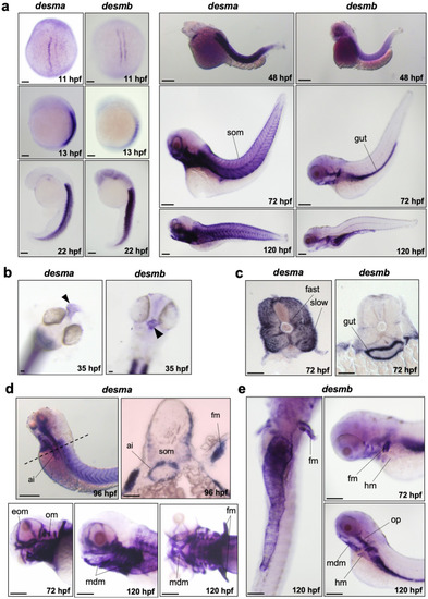
Whole mount in situ mRNA hybridisation of embryos at the indicated stages for antisense probes to desma and desmb. (a) Lateral views are anterior to top, dorsal to left for 13–22 hpf; anterior to left except 11 hpf which is a dorsal view. 48–120 hpf whole mounts are anterior to left, dorsal to top. Scale bar: 250 µm. (b) Arrows indicate heart in frontal view for left panel (desma) and ventral view for right panel (desmb) of 35 hpf embryos. (c) Transversal sections where dorsal is top. (d) Upper left panel is a lateral view at 96 hpf, scale bar: 250 µm. Dashed line represents the position of the transversal section in upper right panel where dorsal is top, scale bar: 100 µm. Lower panels are lateral views of zebrafish heads at 72 and 120 hpf treated with desma probe. Scale bar: 100 µm. (e) Left panel is a ventral view with anterior at top. Right panels are lateral views. Ai, anterior intestine; eom, extraocular muscles; fast, fast muscles; fm, pectoral fin muscles; hm, hypaxial muscles; mdm, mandibular muscles; om, opercular muscles; slow, slow muscles; som, somites. Scale bar: 250 µm.

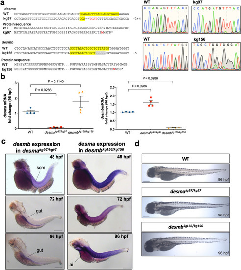
Generation of desma and desmb knockout lines. (a) Alignments and chromatograms wild-type DNA sequences with mutant alleles, and predicted mutant polypeptide sequences. In DNA sequences, yellow highlights gRNA target sequence, hyphens show deleted bases, inserted bases are indicated in red font. In protein sequences, the first residue affected by the frameshift is indicated in red font, asterisks represent early stop codons. Chromatograms were created in SnapGene software (from Insightful Science; available at snapgene.com). (b) Quantitative real-time PCR results showing the expression of desma and desmb mRNA in wild-type and homozygous mutant 96 hpf embryos (N = 4, Mann–Whitney U). (c) Left panel shows whole mount in situ mRNA hybridisation of desmakg97 homozygous embryos at the indicated stages for antisense probes to desmb. Right panel shows whole mount in situ mRNA hybridisation of desmbkg156 homozygous embryos at the indicated stages for antisense probes to desma. Scale bar: 250 µm. Ai, anterior intestine; som, somite. (d) Brightfield pictures of 96 hpf WT and mutant embryos. Scale bar: 250 µm.

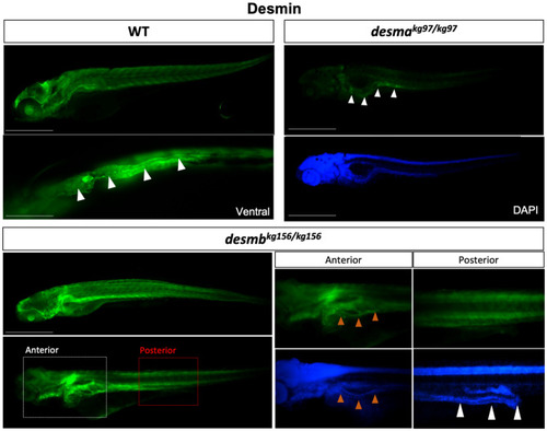
Desmin protein expression in 96 hpf embryos. Whole mount immunofluorescence staining of desmin with anti-desmin polyclonal antibody recognizing both Desma and Desmb, in wild-type and mutant 96 hpf embryos. Left upper panel is lateral view of a WT embryo with anterior to left, dorsal to top, beneath is ventral view with anterior to left, white arrowheads indicate the gut. Right upper panels are lateral views of desmakg97/kg97 with anterior to left, dorsal to top. Lower panel shows desmbkg156/kg156 embryos with anterior to left, dorsal to top. Scale bar: 500 µm. Boxes with dashed lines delineate areas that were zoomed and focused on anterior and posterior segments of the intestine as shown in lower right panels, orange arrowheads indicate anterior intestine, white arrowheads show middle and posterior segments of the intestine.

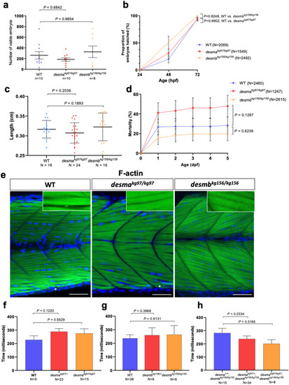
Evaluation of neuromuscular defect in mutant larvae. (a) The number of viable embryos after successful mating of WT fish (n = 10) compared to homozygous desmakg97 (n = 10) (P = 0.6842, Mann–Whitney U) or homozygous desmbkg156 (n = 8) (P = 0.9654, Mann–Whitney U) fish. (b) Comparison of hatching rate between homozygous mutants and WT 24, 48 and 72 hpf embryos At 72 hpf, no significant difference in the hatching rate was found between mutants and WT (desmakg97/kg97 vs. WT, P = 0.9902; desmbkg156/kg156 vs WT, P = 0.3186, repeated measures two-way ANOVA, Bonferroni post hoc test) (c) Body length of 96 hpf WT (N = 18) and homozygous desmakg97 (N = 25) or homozygous desmbkg156 (N = 16) mutant embryos (P = 0.2036 for WT vs. desmakg97/kg97; P = 0.1893 for WT vs. desmbkg156/kg156, Mann–Whitney U). (d) Cumulative mortality rate from 1 to 5 dpf homozygous desmakg97 mutants (N = 1247) compared to WT (N = 2460) (P = 0.1287, repeated measures two-way ANOVA, Bonferroni post hoc test) and homozygous desmbkg156 mutants (N = 2615) compared to WT (P = 0.6239, repeated measures two-way ANOVA, Bonferroni post hoc test). (e) Optical sections of the mid-trunk region of WT and homozygous mutants 96 hpf embryos (N = 8) stained with rhodamine phalloidin (F-actin) with insets showing striations (asterisks indicate occasional wavy fibres in both WT and mutants). Scale bar: 50 µm. (f–h) Touch-evoked escape time (ms) of 48 hpf embryos. Motility experiments were performed blind on siblings from (f) desmakg97/+, (g) desmbkg156/+ or (h) desmakg97/+;desmbkg156/kg156 in-crosses followed by post hoc genotyping. (f) Heterozygous desmakg97 embryos (N = 23) were compared to WT (N = 9) (P = 0.122, Mann–Whitney U) and desmakg97/kg97 (N = 15) compared to WT (P = 0.5529, Mann–Whitney U). (g) Heterozygous desmbkg156 embryos (N = 6) were compared to WT (N = 38) (P = 0.3968, Mann–Whitney U) and desmbkg156/kg156 (N = 6) compared to WT (P = 0.6131, Mann–Whitney U). (h) Homozygous double mutants (N = 8) were compared to desma+/+;desmbkg156/kg156 (n = 15) (P = 0.3165, Mann–Whitney U) and desmakg97/+;desmbkg156/kg156 embryos (n = 34) compared to desma+/+;desmbkg156/kg156 (P = 0.2334, Mann–Whitney U).

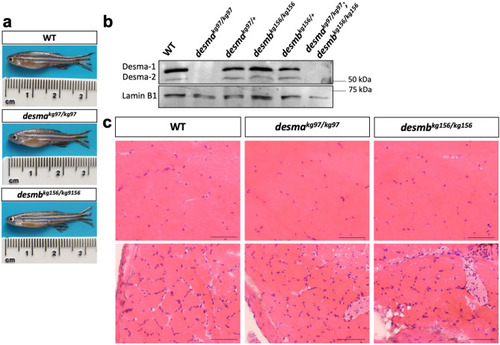
Desmin protein expression and histological examination of adult skeletal muscle. (a) Photographs of 1 year-old anesthetized fish. (b) Immunoblotting of skeletal muscle protein extracts of WT, desmakg97/kg97, desmakg97/+, desmbkg156/kg156, desmbkg156/+ and desmakg97/kg97;desmbkg156/kg156 double mutant fish probed with anti-desmin antibody. Lamin B1 was labeled on the same blot as loading control. Uncropped full-length pictures of the blot is available in Supplementary Figure S5 online. (c) Transversal cryosections of mid-trunk somites from two different areas of WT and homozygous mutant adults stained with hematoxylin eosin showing no pathological changes. Scale bar: 25 µm.

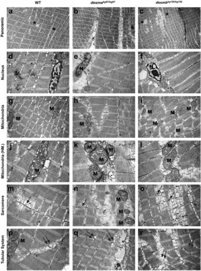
Ultrastructural features of adult WT and mutant skeletal muscle tissue. First, second and third columns present adult WT, desmakg97/kg97, desmbkg156/kg156 skeletal muscle tissues’ electron micrographs, respectively. First row indicates the generally aligned sarcomeric structures with the presence of seldom disruptions (ac, asterisks). Second row shows centrally (d) and peripherally (e,f) located healthy nuclei (N) with peripherally condensed chromatin and well-defined nucleoli. Third and fourth row (g–l) shows pleomorphic mitochondria (M) with well-defined tubular cristae. Fifth and sixth rows present the Z line streaming (arrows in mo) and enlargement (mr) of the T-tubule systems related to the triads (double arrows). Uranyl acetate, lead citrate. HM high magnification. Magnifications are as follows: (ac) 8000 ×, (d) 25,000 ×, (e,f) 20,000 ×, (gi) 15,000 ×, (j) 40,000 ×, (k) 60,000 ×, (l) 30,000 ×, (mo) 20,000 ×, (p) 25,000 × and (q,r) 20,000 ×.

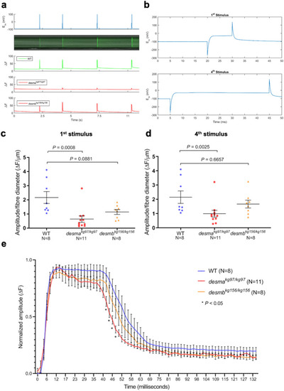
Calcium flux in isolated fibres. (a) Calcium flux along fibres was monitored in vivo by Fluo-4 AM after four consecutive depolarizing stimuli. Top panel, membrane potential responses to current stimulus pulses with varying durations of 10, 20, 30, 40 ms with the amplitude of the current pulse kept constant at 100 mV and the duration increased by 10 ms at each pulse. Middle panel, evoked calcium transients in line scan mode (tx) to current stimuli. Bottom panel, integrated emission signal as a function of time for each experimental group. (b) Membrane potential response to the first stimulus of 10 ms (top) or to the fourth stimulus of 40 ms. (c, d) Baseline corrected amplitude divided by fibre diameter (ΔF/µm) of the calcium emission signals during the first (10 ms) stimulus and fourth (40 ms) stimulus were compared between mutants and WT. (e) Time course analysis of the baseline corrected and normalized amplitude of the calcium transient from the longest stimulus (40 ms) was represented as mean amplitude values as a function of time. Homozygous desmakg97 fibres (N = 11, red curve) or homozygous desmbkg156 fibres (N = 8, orange curve) were compared to WT (N = 8, blue curve). (* indicates time points where P < 0.05, repeated measures two-way ANOVA, Bonferroni post hoc test).

Acknowledgments
This image is the copyrighted work of the attributed author or publisher, and ZFIN has permission only to display this image to its users. Additional permissions should be obtained from the applicable author or publisher of the image. Full text @ Sci. Rep.