FIGURE SUMMARY
Title

10-Gingerol Suppresses Osteoclastogenesis in RAW264.7 Cells and Zebrafish Osteoporotic Scales

Authors
Zang, L., Kagotani, K., Nakayama, H., Bhagat, J., Fujimoto, Y., Hayashi, A., Sono, R., Katsuzaki, H., Nishimura, N., Shimada, Y.
Source
Full text @ Front Cell Dev Biol

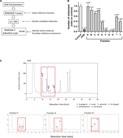
Ginger hexane extract (GHE) fractions containing gingerols suppress osteoclastogenesis in RAW264.7 cells. (A) Experimental design to identify anti-osteoclastic constituents in GHE. (B) The number of TRAP-positive multinucleated osteoclasts containing more than 11 nuclei. n = 3, error bars indicate SE; p values are calculated compared to control. The representative TRAP stained images for each fraction are shown in Supplementary Figure 1. (C) Chromatograms of HPLC analysis of GHE and Fractions F, G, and H. The peaks were detected at UV 228 nm absorbance. The peak numbers 1, 2, 3, 4, 5, and 6 refer to 6-gingerol, neral, citral, 6-shogaol, phellandrene, and farnesene, respectively. Identification of P1, P2, and P3 indicated by arrowheads was performed by LC/MS analysis.

10-gingerol suppresses osteoclastogenesis in RAW264.7 cells. (A) Representative images of sRANKL-treated RAW264.7 cells with 6-, 8-, or 10-gingerol. Each treatment was carried out at 2.5 μM. Red indicates the TRAP-stained area. The lower panels show the magnified images of sRANKL-treated RAW264.7 cells with or without gingerols. (B) Quantitative analysis of TRAP-positive multinucleated osteoclasts of (A). n = 8, error bars indicate SE; p values are calculated compared to vehicle with sRANKL. (C) Representative images for the pit formation assay with or without 10-gingerol. White indicates pits. (D) Bone resorption activities of panel (C) were quantified by the released FACS from the well bottom. n = 3, error bars indicate SE; p < 0.05 vs. vehicle with sRANKL.

10-gingerol suppresses osteoclastogenesis in zebrafish osteoclastic scales. (A) Representative images for prednisolone (PN) treated regenerating zebrafish scales with 10-gingerol, GHE, or alendronate (AL; positive control). The lower panels show magnified images of the right lateral regions of the upper scales. Red color indicates the TRAP-positive area. The dark area in PN indicates the concentration of TRAP-positive cells. The black arrow indicates the resorbed edges, and white arrows show the resorption pits. For the experimental design of the zebrafish study, please see Supplementary Figure 2. (B) Quantitative analysis of (A). Integrative densities of the red areas were measured using ImageJ. n = 15–20, error bars indicate SE; ∗∗p < 0.01 vs. PN. (C) Scales after calcein and alizarin red S staining. The upper panels show calcein staining, Alizarin red staining, and merged images, respectively. Scale bar = 500 μm.

Gene expression analysis of RAW264.7 cells and zebrafish scales treated with 10-gingerol. (A,B) qPCR analysis of osteoclast marker genes in RAW264.7 cells with 10-gingerol. Oscar: Osteoclast-associated immunoglobulin-like receptor; Dc-stamp: Dendrocyte expressed seven transmembrane protein; Traf6: Tumor necrosis factor receptor-associated factor 6; Ctsk: Cathepsin k; Trap: Tartrate-resistant acid phosphatase; Mmp9: matrix metalloproteinases-9; Nfatc1: nuclear factor of activated T cells cytoplasmic 1. n = 3, error bars indicate SE; *p < 0.05, **p < 0.01. The data for GHE treatment were referred from our previous study (Ito et al., 2016). nd indicates no data. (C) qPCR analysis of osteoclast marker genes in osteoporotic zebrafish scales with 10-gingerol, GHE, or AL. ctsk: cathepsin k; mmp2: matrix metalloproteinases-2. *p < 0.05, **p < 0.01. n = 5, error bars indicate SE. (D) Fluorescent immunohistochemistry for Nfatc1 in RAW264.7 cells treated with sRANKL for 3 days. Red indicates NFATc1, and blue indicates nuclei (DAPI). (E) qPCR analysis of Nf-kb1 and Nf-kb2 in RAW264.7 cells with 10-gingerol. Nf-kb1: Nuclear factor kappa B subunit 1; Nf-kb2: Nuclear factor kappa B subunit 2. *p < 0.05. n = 5, error bars indicate SE. (F,G) qPCR analysis of nf-kb1, nf-kb2, (F) and rankl(G) in osteoporotic zebrafish scales (PN) with 10-gingerol, GHE, or AL. rankl: receptor activator of nuclear factor kappa B ligand. #p < 0.1, *p < 0.05 vs. PN group. n = 5, error bars indicate SE. (H) Cell-free CTSK inhibitor assay of 10-gingerol (100 μM) and GHE (100 μg/mL). FF-FMK (10 μM) was used as a positive control (98.3% inhibition to the negative control). **p < 0.01 vs. negative control. n = 3, error bars indicate SE.

Acknowledgments
This image is the copyrighted work of the attributed author or publisher, and ZFIN has permission only to display this image to its users. Additional permissions should be obtained from the applicable author or publisher of the image. Full text @ Front Cell Dev Biol