FIGURE SUMMARY
Title

Specific fibroblast subpopulations and neuronal structures provide local sources of Vegfc-processing components during zebrafish lymphangiogenesis

Authors
Wang, G., Muhl, L., Padberg, Y., Dupont, L., Peterson-Maduro, J., Stehling, M., le Noble, F., Colige, A., Betsholtz, C., Schulte-Merker, S., van Impel, A.
Source
Full text @ Nat. Commun.

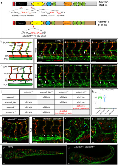
Lymphangiogenesis is completely abolished in <italic>adamts3</italic>; <italic>adamts14</italic> double mutant embryos.

a Schematic of Adamts3 and Adamts14 protein structures depicting the predicted effects of the indicated deletion alleles. Shown are the aa positions of the deletion-induced frame shifts (red letters) as well as the position of the resulting premature stop codons. SP signal peptide, MPD metallopeptidase domain, ACR ADAM cysteine-rich domain 2, TSP1 thrombospondin type-1. b Schematic representation of the wild-type trunk vasculature at 48 hpf. ce, gi, and lqflt4:mCitrine; flt1:tdTomato double transgenic embryos highlighting arterial ECs in red and venous and lymphatic structures in green. In wild-type (c), adamts3 (d) or adamts14 single mutants (e), PL cells align at the HM at 48 hpf (arrows). f Schematic representation of the trunk vasculature at 5 dpf with the thoracic duct (TD) being located between DA and PCV. Compared with wild type (g), neither homozygous adamts3 (h) nor adamts14 mutants (i) exhibit lymphatic defects at 5 dpf (arrows). j Table summarizing the analysis of lymphatic phenotypes in all double mutant combinations for adamts2, adamts2_like, adamts3, and adamts14. k At 48 hpf, the number of PLs was quantified in n = 53 siblings and n = 30 adamts3; adamts14 double mutants and the number of vISVs was quantified in n = 37 siblings and n = 11 double mutants. Box-and-whisker plots show median and quartiles with whiskers indicating minimum/maximum values (source data are provided as a Source Data file). ladamts3; adamts14 double mutants lack PLs (white asterisks) and do not form vISVs (blue asterisks) at 48 hpf. m At 5 dpf, the TD is completely absent in double mutants (white asterisks). Facial lymphatics (blue arrows) and meningeal lymphatics (yellow arrow) are formed in siblings at 5 dpf (n) but are absent in adamts3; adamts14 double mutants (o). In contrast to their siblings (p), double mutants (q) display a variably strong curvature of the trunk. Scale bars in ce, gi, lo: 50 µm; p, q: 100 µm. aa amino acid, bp base pair, dpf days post fertilization, ECs endothelial cells, DA dorsal aorta, DLAV dorsal longitudinal anastomotic vessel, hpf hours post fertilization, HM horizontal myoseptum, a/vISV arterial/venous intersegmental vessel, PCV posterior cardinal vein, PL parachordal lymphangioblast, VTA vertebral artery.

Adamts3 and Adamts14 are both able to process pro-Vegfc.

a Schematic depiction of the in vivo Vegfc-processing assay. In wild-type embryos, floorplate expression of full-length Vegfc results in venous hyper-branching at 48 hpf. beflt4:mCitrine; flt1:tdTomato transgenic embryos with arterial structures in red and venous and lymphatic structures in green. b, c While forced expression of full-length zebrafish Vegfc from the floorplate leads to hyper-branching in wild-type embryos (b, arrows), this dominant phenotype is not visible in adamts3; adamts14 double mutants (c, asterisks). Allelic combinations with one remaining wild-type copy of either adamts3 (d) or adamts14 (e) develop the Vegfc-induced hyper-branching phenotype. Blue lines indicate the floorplate position (n = 225 analyzed embryos). fi Mosaic expression of full-length human VEGFC in floorplate cells (marked by tagRFP expression) results in local venous hyper-branching in wild-type embryos (f), but not in adamts3;adamts14 double mutants (g). One functional copy of either gene (h, i) is sufficient to restore the dominant phenotypes (n = 157 injected tagRFP+ embryos). Scale bars: 50 µm. DA dorsal aorta, DLAV dorsal longitudinal anastomotic vessel, hpf hours post fertilization, HM horizontal myoseptum, a/vISV arterial/venous intersegmental vessel, PCV posterior cardinal vein, PL parachordal lymphangioblast. j N-terminal processing of human VEGFC by ADAMTS3 and ADAMTS14 proteases. Conditioned medium from HEK293 cells expressing full-length VEGFC was incubated with ADAMTS3, ADAMTS14, or control buffer, in the presence or absence of EDTA. VEGFC forms were analyzed by Western blotting. In absence of active ADAMTS proteins (lanes 2, 4, and 5), VEGFC can be detected as a 58 kDa (full-length VEGFC without SP) and a 31 kDa form (C-terminally processed VEGFC without SP). In the presence of active ADAMTS14 (lane 1), the 58 kDa form is converted into a 45 kDa polypeptide while the 31 kDa form is processed into the fully mature 21 kDa VEGFC, which is in line with N-terminal processing of VEGFC proteins. A similar shift in VEGFC forms is detectable in the presence of active ADAMTS3 (lane 3) [source data are provided as a Source Data file]. Schematic depicting different VEGFC forms and their molecular weights. SP signal peptide, NT N-terminal propeptide, VHD VEGF homology domain, CT C-terminal propeptide.

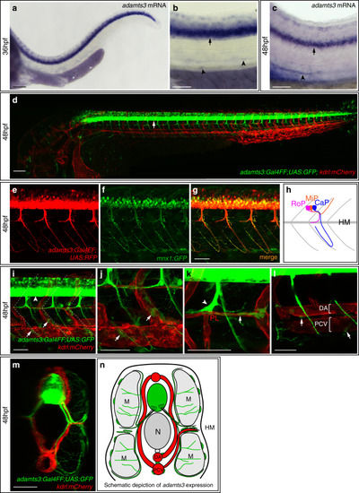
<italic>adamts3</italic> is expressed by motoneurons and additional cell types during venous sprouting stages.

ac Whole mount in situ hybridization against adamts3 at 36 and 48 hpf indicates strong expression in the spinal cord (arrow) and weaker expression of adamts3 transcripts around the axial blood vessels in the trunk (arrowheads). d The adamts3:Gal4FF; UAS:GFP transgenic reporter line shows very prominent expression (in green) in neuronal cells (arrow). eg The adamts3:Gal4FF; UAS:RFP transgene shows expression in trunk neurons and axons that are also labeled by the motoneuronal marker mnx1:GFP at 48 hpf. h Diagram of the three different types of motoneurons per segment, differing in their axonal projections which either extend along the HM region (rostral primary motoneuron, RoP, shown in magenta), toward the dorsal side of the trunk (middle primary motoneuron, MiP, highlighted in orange) or that extend further ventrally, passing by the main axial blood vessels (caudal primary motoneuron, CaP shown in blue). iladamts3:Gal4FF; UAS:GFP; kdrl:mCherry transgenic embryos, highlighting adamts3 expression domains in green and ECs in red at 48 hpf. i Higher magnification of the trunk region, showing adamts3 reporter expression not only in neurons (arrowhead) but also in non-neuronal cells located at the segment boundaries (arrows). White lines indicate the position of the segment boundaries. j Higher magnification of the ventral trunk region, indicating the position of adamts3-expressing cells along the segment boundaries (arrows). k Partial z-projection of the HM region indicating expression of the adamts3 reporter (in green) in the RoP axon (arrowhead) and in cells along the HM (arrow), which are in close proximity to the PL cells (in red). l In partial z-projections of the trunk, individual adamts3-positive cells around the DA and the PCV are apparent (arrows). m Virtual cross section of the trunk region shown in i. n Schematic cross view illustrating the different adamts3 expression domains (green) in the trunk.; Scale bar in a, d: 100 µm; b, c, eg, im: 50 µm. DA dorsal aorta, ECs endothelial cells, hpf hours post fertilization, HM horizontal myoseptum, M muscle, N notochord, PCV posterior cardinal vein, PL parachordal lymphangioblast.

<italic>adamts14</italic> expression can be detected in the floorplate and at the horizontal myoseptum.

al Whole mount in situ hybridization for adamts14. ad At 26 hpf, adamts14 transcripts are detectable in the floorplate (black arrow), in cells along the ventral aspect of the segment boundaries (white arrow) and in cells at the HM (black arrowhead). d Zoom-in of the squared region in c. eh At 36 hpf, adamts14 expression is visible in the floorplate (black arrow), in cells located at the level of the axial blood vessels (white arrow) and in cells at the HM (black arrowhead). h Zoom-in of the indicated region in g. il Expression of adamts14 is still apparent in the floorplate (black arrow) at 48 hpf as well as in cells at the HM (black arrowhead). l Enlargement of the boxed region in k. m, nadamts14 RNA granules can be detected in cells located in the HM region by RNAscope at 48 hpf. n Overlay of the bright field and confocal image for the indicated region in m, showing adamts14 RNA granules in green. o Schematic cross view of the trunk illustrating the adamts14 expression domains (yellow). Scale bars in a, e, i: 100 µm; b, c, f, g, j, k, m: 50 µm. DA dorsal aorta, hpf hours post fertilization, HM horizontal myoseptum, M muscle, N notochord, PCV posterior cardinal vein, PL parachordal lymphangioblast.

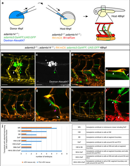
<italic>adamts3</italic> expression via motoneurons or mesenchymal cells can rescue venous sprouting and PL formation in <italic>adamts3</italic>; <italic>adamts14</italic> double mutants.

a Diagram depicting the heterochronic transplantation approach: Dextran-Alexa647 is injected into one-cell stage adamts14−/−; adamts3:Gal4FF; UAS:GFP embryos (donor embryos). Cells are transferred from donor embryos to adamts3; adamts14 double mutants at 6 hpf (host embryo) and the effects are assessed at 48 hpf. b, c In cases where transplanted cells contributed to adamts3-expressing motoneuron clusters (green arrowhead), venous ISVs (white arrowhead), and PL cells (white arrow) formed in double mutants. c Transplanted cells labeled by Dextran-Alexa647. d When transplants gave rise to adamts3+ cells at three locations, the HM, the ventral segment boundaries, and close to the DA and PCV, a local rescue of the formation of PL cells and intersegmental veins in adamts3; adamts14 double mutant embryos was visible. e Zoom-in of the boxed region in (d) with adamts3-expressing cells at the segment boundary highlighted by an arrow and transplanted cells at the HM marked by an arrowhead. f, g Transplantation of cells contributing to adamts3-expressing cells at the ventral segment boundaries (blue arrow) and to cells located close to the DA (magenta arrow) and the PCV (white arrow) also resulted in a locally restricted rescue of PL and vISV formation. g Zoom-in of the indicated region in f. h, i Cases, in which the transplants gave rise to adamts3+ cells at the HM (arrowhead in i) and the region around the PCV (arrow in i), but not to the cells along the ventral segment boundaries showed a rescue of PL cells and vISVs. j Overview about the results obtained from 23 rounds of cell transplantations. Each round contained 144 recipient embryos obtained from an adamts3+/−; adamts14−/− incross of which a quarter was expected to be double mutant (in total 828). Embryos showing a rescue of PL or vISV development were selected for imaging and genotyping. All embryos shown in (j) are adamts3; adamts14 double mutants. Scale bars: 50 µm. DA dorsal aorta, hpf hours post fertilization, PCV posterior cardinal vein, PL parachordal lymphangioblast, vISV venous intersegmental vessel.

<italic>adamts14</italic>-expressing floorplate and mesenchymal cells can rescue venous sprouting and PL formation in <italic>adamts3</italic>; <italic>adamts14</italic> double mutants.

a Scheme outlining the transplantation assay: embryos homozygous mutant for adamts3 were injected with Dextran-Alexa647 to mark the donor cells. At 4 hpf, cells from the donor were transplanted into adamts3; adamts14 double mutant host embryos (6 hpf). Host embryos were subsequently analyzed for rescue of venous sprouting and the location of the transplanted material at 48 hpf. bd Transplantation of cells contributing to the floorplate (arrow) resulted in the formation of intersegmental veins and venous sprouts reaching the HM region. c Dextran-Alexa647 labeled transplanted tissue. d Virtual cross section showing the transplanted floorplate cells (white) in the rescued area. eh In cases where the transplanted cells contributed to muscle but also to individual cells at the HM (magenta arrow in f, g), at segment boundaries (blue arrow in f) and also to cells located directly adjacent to the DA and PCV (blue arrow in h), PL formation and vISV formation was locally restored in adamts3; adamts14 double mutants. f Dextran-Alexa647 channel highlighting the transplanted tissue. g, h Partial z-projections of the boxed regions in (e) indicating transplanted cells in white. ik Transplants giving only rise to muscle tissue did not rescue the venous sprouting defects in double mutants. j Dextran-Alexa647 labeled transplanted tissue. k Zoom-in of the boxed HM region in j. ln Cell transplantations giving rise to tissues including cells located at the HM (arrow in m) resulted in a rescue of PL and vISV formation in this area at 48 hpf. n Virtual cross section indicating the position of the transplanted cell at the HM (arrow). o Graph summarizing the results from 13 rounds of cell transplantations. Each round contained 144 recipient embryos derived from an adamts3+/−; adamts14−/− incross. From the expected 468 double mutant recipients, embryos showing PL or vISV rescue were selected for imaging and genotyping. All embryos shown in (o) are adamts3; adamts14 double mutants. Scale bars: 50 µm. DA dorsal aorta, HM horizontal myoseptum, hpf hours post fertilization, PCV posterior cardinal vein, PL parachordal lymphangioblast, vISV venous intersegmental vessel.

Non-neuronal <italic>adamts3</italic>-expressing cells co-express <italic>vegfc</italic> and <italic>ccbe1</italic>.

a Strategy for transcript analysis of adamts3-expressing cell populations by RNA sequencing. adamts3:Gal4FF;UAS:GFP; NBT:dsRed-positive trunks were dissociated at 48 hpf and cells were FAC-sorted into (1) adamts3-expressing neurons (GFP + /dsRed+, yellow arrowhead), (2) adamts3 + non-neuronal cells (GFP+, green arrowheads) and (3) a negative control. Pooled cell populations were analyzed by RNA sequencing. b Summary of RNA sequencing analysis. Values reflect fold-changes in transcript abundance for indicated genes upon comparison of cell populations stated in the column headings. To identify significant gene expression differences, p values were determined by Wald test and an FDR < 0.05 and FDR-adjusted p value < 0.1 cutoff was used (source data are provided as a Source Data file). c Lateral view of a vegfc:Gal4FF; UAS:GFP; kdrl:mCherry transgenic embryo at 48 hpf. d Higher magnification showing vegfc expression in HM cells (white arrowhead), in the hypochord (blue arrowhead), the DA (yellow arrowhead), in arteries (green arrowhead) and in neurons (magenta arrowhead). e Zoom-in of the boxed region in (d) highlighting vegfc-expressing cells at the HM (arrowhead). f Virtual cross section focusing on the HM, showing juxtaposition between a PL and vegfc-expressing cells (arrowhead). g Lateral view of a ccbe1:mCitrine; kdrl:mCherry transgenic embryo at 48 hpf showing ccbe1 expression in the pronephros (blue arrow) and epiphysis (white arrow). h Higher magnification demonstrating ccbe1 expression at the HM (arrowhead). i Partial projection of the marked region in (h) showing ccbe1-expressing cells at the HM (white arrowhead) and the segment boundary (yellow arrowhead). j Virtual cross section indicating ccbe1+ cells at the HM (arrowhead). k, m, o Lateral views (partial z-projections) and l, n, p virtual cross sections of the HM region at 48 hpf. Arrowheads indicate cells with co-expression of the indicated transgenes. k, ladamts3 and vegfc are co-expressed by individual cells at the HM. m, n Mesenchymal cells at the HM co-express adamts3 and ccbe1. o, p Overlapping expression of vegfc and ccbe1 in HM cells. Scale bars in a, d, h: 50 µm; c, g: 100 µm; f, j, k, m, o 25 µm; l, n, p: 10 µm. DA dorsal aorta, HM horizontal myoseptum, hpf hours post fertilization, neg. negative cell population, sig. significant.

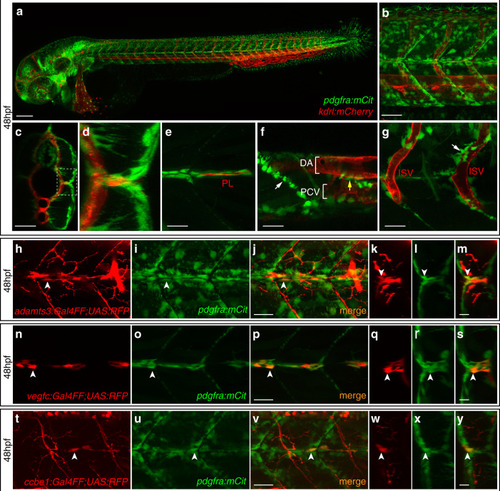
Co-expression of <italic>adamts3</italic>, <italic>ccbe1</italic>, and <italic>vegfc</italic> with <italic>pdgfra</italic> at the horizontal myoseptum.

ag Confocal images of pdgfra:mCitrine; kdrl:mCherry double transgenic embryos at 48 hpf. a Lateral view giving an overview of the pdgfra-expression domains. b Higher magnification of the trunk region above the yolk extension. c, d Virtual cross section of the trunk shown in b. d Zoom-in of the boxed area in (c) showing the HM region in detail. eg Partial projections of (b). e At the HM, pdgfra-expressing cells (in green) are surrounding the PL cells (in red). fpdgfra is expressed by cells located along the segment boundaries (white arrow) as well as by individual cells located around the main axial blood vessels (yellow arrow). g At the level of the ISVs, individual pdgfra+ cells locate in close proximity to the blood vessels. hj, np, and tv Lateral views and km, qs, and wy virtual cross sections of the HM region in double transgenic embryos at 48 hpf. White arrows highlight cells with co-expression. hm A subpopulation of pdgfra-expressing cells (green) is co-labeled by the adamts3 reporter transgene (red) at the HM. ns Cells at the HM co-express vegfc (red) and pdgfra (green). ty All ccbe1-expressing cells at the HM (red) show co-expression of pdgfra (green). Scale bar in a: 100 µm; b, c: 50 µm; ej, np, tv: 25 µm; km, qs, wy: 10 µm. DA dorsal aorta, HM horizontal myoseptum, hpf hours post fertilization, ISV intersegmental vessel, PCV posterior cardinal vein, PL parachordal lymphangioblast.

Single-cell RNA sequencing identifies fibroblast subpopulations expressing <italic>adamst3, adamts14, ccbe1</italic>, <italic>and vegfc</italic>.

a Scheme of the partial dissociation approach and the work flow. Note that embryonic trunks were not completely dissociated in order to obtain a higher proportion of cells from more superficial tissues while reducing the amount of cells in the suspension originating from deeper tissues. b Pagoda2 clustering of the individual cell transcriptomes gives rise to 24 clusters (labeled as C1-C24). cn t-SNE plots indicating the transcript levels of the respective gene in each sequenced cell. Expression of the general fibroblast markers pdgfra (c), col1a1a (d), lum (e), and dcn (f). Expression of ccbe1 (g) and vegfc (h) is largely restricted to cells within cluster 2 as is the case for the expression of the genes en1a (i) and en1b (j). adamts3- (k) and adamts14-expressing cells (l) are distributed over several clusters. m, n While cxcl12a-expressing cells are enriched in cluster 2, svep1-expressing cells can be found within several different clusters. t-SNE plots highlighting cells co-expressing vegfc and ccbe1 (o), adamts3 and vegfc (p), or adamts3 and ccbe1 (q) as well as cells co-expressing adamts14 and vegfc (r) or adamts14 and ccbe1 (s). Symbol shapes in the t-SNE plots indicate from which FACS run the cells had been obtained (circles and triangles originate from sorts using vegfc:Gal4FF; UAS:GFP; kdrl:mCherry embryos, cells indicated by squares stem from the FAC-sorting of pdgfra:mCitrine embryos), color scales reflect the relative transcript levels. hpf hours post fertilization.

Local Vegfc processing at the HM is required to guide endothelial cells to this region.

a Schematic depiction of the mature human VEGFC overexpression construct and the vegfc:Gal4FF transgenic line, which was used to drive mosaic expression of the construct upon transient injection. bqvegfc:Gal4FF positive embryos were injected with the overexpression construct at the one-cell stage and imaged at 48 hpf. All embryos shown are adamts3; adamts14 double mutant, transgenic for flt4:mCitrine (green) and express UAS:ΔNΔC-VEGFC-P2A-RFP (red). b, c Expression of UAS:ΔNΔC-VEGFC-P2A-RFP in hypochord cells (white arrow) rescues vein formation. d, e Local vISV rescue upon expression of mature VEGFC in DA cells (white arrowhead). fi Venous hyper-sprouting with ectopic venous structures formed in the vicinity of hypochord cells (f, g, arrows) or DA cells (h, i, arrowheads) with expression of the ΔNΔC-VEGFC construct. j, kUAS:ΔNΔC-VEGFC-P2A-RFP positive fibroblasts recruit PL cells to the HM region. l, m Expression of mature VEGFC in hypochord cells and in fibroblasts at the HM rescues vISV and PL formation in adamts3; adamts14 double mutant embryos. Note the close proximity of the PL cell to the ΔNΔC-VEGFC producing fibroblasts. n, o Combined expression of mature VEGFC in DA cells and fibroblasts at the HM results in the formation of vISV and PL cells close by. p, q Simultaneous expression of ΔNΔC-VEGFC in hypochord cells and in fibroblasts at the HM results in a local rescue of PL and vISV formation but can additionally lead to a hyper-branching of venous structures at the level of the DA. c, e, g, i, k, m, o, q: Zoom-in image of the indicated regions in b, d, f, h, j, l, n, p. Scale bars: 50 µm. DA dorsal aorta, HM horizontal myoseptum, hpf hours post fertilization, PL parachordal lymphangioblast, vISV venous intersegmental vessel.

In situ hybridization against vegfc using a milder proteinaseK treatment reveals expression at the horizontal myoseptum. a, b) vegfc transcripts were detected in the hypochord, dorsal aorta (arrow) and in developing intersegmental arteries at 26hpf. b) Higher magnification of the trunk region with prominent staining of the hypochord (arrow). c, d) At 36hpf vegfc transcripts were detected in cells at the HM (black arrow), the hypochord and the dorsal aorta (orange arrow). d) Higher magnification of the trunk region. e, f) Expression of vegfc within the DA and in cells at the HM persist at 48hpf. f) High magnification of the trunk focusing at the HM level. Scale bars in a, c, e: 100μm; b, d, f: 50μm. hpf: hours post fertilization, DA: dorsal aorta, HM: horizontal myoseptum.

EXPRESSION / LABELING:
Gene:
Fish:
Anatomical Terms:
Stage Range: Prim-5 to Long-pec

Fibroblasts at the horizontal myoseptum express Engrailed proteins. a-i) HM region of 48hpf embryos that were stained with anti-Engrailed (red) and anti-GFP (green) antibodies. a-c) All cells expressing the vegfc:Gal4FF; UAS: GFP reporter at the HM do co-express Engrailed proteins. d-f) fpdg-fra:mCitrine positive fibroblasts at the HM are also positive for Engrailed. g-i) Partial z-projections of the HM region reveal a co-expression of the adamts3 reporter and Engrailed proteins within fibroblasts. j, m) Two additional examples of genes whose transcripts are highly enriched within fibroblast cluster 2 are nuak1b and tgbl1 (see Supplementary Tables 1-3). The mRNA of both genes (k, l, n, o) can be detected specifically at the midline by ISH at 48hpf, thereby validating the notion that the cells in cluster 2 represent the fibroblast subpopulation located at the HM. Scale bars in a-i: 25 μm; k, l, n and o: 50 μm. hpf: hours post fertilization, HM: horizontal myoseptum

EXPRESSION / LABELING:
Genes:
Fish:
Anatomical Term:
Stage: Long-pec
Acknowledgments
This image is the copyrighted work of the attributed author or publisher, and ZFIN has permission only to display this image to its users. Additional permissions should be obtained from the applicable author or publisher of the image. Full text @ Nat. Commun.