FIGURE SUMMARY
Title

Functional analysis of new human Bardet-Biedl syndrome loci specific variants in the zebrafish model

Authors
Castro-Sánchez, S., Suarez-Bregua, P., Novas, R., Álvarez-Satta, M., Badano, J.L., Rotllant, J., Valverde, D.
Source
Full text @ Sci. Rep.

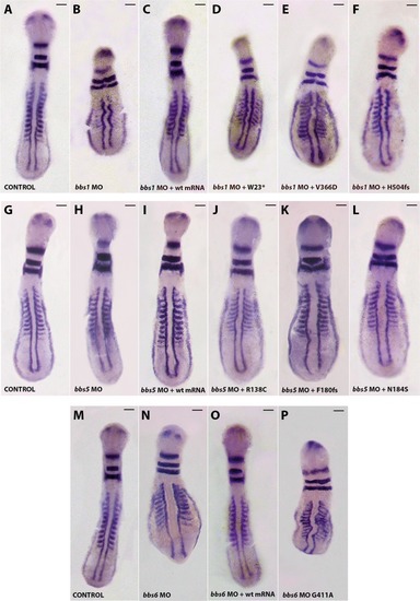
Phenotypes of zebrafish embryos at 8–12 ss, after whole mount in situ hybridization. Knockdown of zebrafish bbs1 (AF), bbs5 (G–L) and bbs6 (MP) genes affects body axis/length, notochord and somite morphology. Morphology of the controls (A,G,M; dorsal view anterior to the top), morpholino (B,H,M; dorsal view anterior to the top), morpholino plus WT human BBS capped-mRNA (C,I,O), and morpholino plus sense capped-mRNA of different human BSS variants (DF,JL,P; dorsal view anterior to the top) zebrafish at 12 somites (ss). The embryos were fixed for in situ hybridization of myoD/krox20/pax2. Injection of WT human BSS1, BSS5 and BSS6 capped-mRNA rescues the phenotype of bbs1, bbs5 and bbs6 MO-injected embryos, respectively. However, the mutated capped-mRNA of the BBS particular changes of each loci (W23*, V366D, H504fs, R138C, F180fs, N184S, G411A – abbreviated nomenclature) were not able to rescue the respective phenotype of BBS1, BBS5 and BBS6 MO-treated embryos. Scales bars: 100 µm.

Representative images of KVs and comparison of average cilia length corresponding to BBS1 conditions. Projections of confocal z-stacks of Alexa fluor 594 and Dapi stained cilia located in Kupffer’s vesicle (KV) from control (A) and bbs MO-injected embryos (B), bbs MO-injected embryos plus WT capped-mRNA (C) and bbs MO-injected embryos plus BBS1 new changes (W23*, V366D, H504fs) capped-mRNA. (DF) Note the reduction of the cilia in bbs1-MO- and BBS1 variants capped-mRNA co-injected embryos. (G) Comparison of average cilia length. *p < 0.01, **p < 0.001. Scales bars: 10 µm.

PHENOTYPE:
Fish:
Knockdown Reagent:
Observed In:
Stage Range: 5-9 somites to 10-13 somites

Representative images of KVs and comparison of average cilia length corresponding to BBS5 conditions. Projections of confocal z-stacks of Alexa fluor 594 and Dapi stained cilia located in Kupffer’s vesicle (KV) from control (A) and bbs5 MO-injected embryos (B), bbs5 MO-injected embryos plus WT capped-mRNA (C) and bbs5 MO-injected embryos plus BBS5 new changes (R138C, F180fs, N184S) capped-mRNA. (DF) Note the reduction of the cilia in bbs5 MO and BBS5 variants R138C and N184S capped-mRNA co-injected embryos. (G) Comparison of average cilia length. *p < 0.01, **p < 0.001. Scales bars: 10 µm.

PHENOTYPE:
Fish:
Knockdown Reagent:
Observed In:
Stage Range: 5-9 somites to 10-13 somites

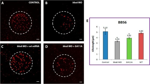
Representative images of KVs and comparison of average cilia length corresponding to BBS6 conditions. Projections of confocal z-stacks of Alexa fluor 594 and Dapi stained cilia located in Kupffer’s vesicle (KV) from control (A) and bbs6 MO-injected embryos (B), bbs6 MO-injected embryos plus WT capped- mRNA (C) and bbs6 MO-injected embryos plus BBS6 new changes (G411A) capped-mRNA. (D) Note the reduction of the cilia in bbs6 MO-injected and BBS6 variant capped-mRNA co-injected embryos. (E) Comparison of average cilia length. *p < 0.01, **p < 0.001. Scales bars: 10 µm.

PHENOTYPE:
Fish:
Knockdown Reagent:
Observed In:
Stage Range: 5-9 somites to 10-13 somites

ZFIN is incorporating published figure images and captions as part of an ongoing project. Figures from some publications have not yet been curated, or are not available for display because of copyright restrictions.

Acknowledgments
This image is the copyrighted work of the attributed author or publisher, and ZFIN has permission only to display this image to its users. Additional permissions should be obtained from the applicable author or publisher of the image. Full text @ Sci. Rep.