FIGURE SUMMARY
Title

TAMM41 is required for heart valve differentiation via regulation of PINK-PARK2 dependent mitophagy

Authors
Yang, R.M., Tao, J., Zhan, M., Yuan, H., Wang, H.H., Chen, S.J., Zhu, C., de Thé, H., Zhou, J., Guo, Y., Zhu, J.
Source
Full text @ Cell Death Differ.

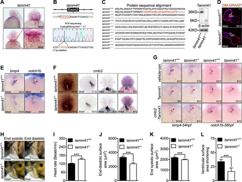
Tamm41 is indispensable for zebrafish cardiogenesis. a WISH analysis of tamm41 expression in zebrafish embryos at 36 hpf and 48 hpf. Red dotted lines delineate how tamm41 expressed. b Schematic diagram of tamm41 targeted crispr9 design, and transmittable F1 carrying 7 bp deletion in the exon2 of tamm41 was generated. c The deletion caused a frame shift and generated a premature stop codon leading to truncated Tamm41 (6KD) generation without most of the functional domains. d Representative images show DR-GRASP staining of 56 hpf tamm41+/+ and tamm41−/− hearts. While several layers of myocardium (upper panel outlined by white dashed line) form adjacent to the endocardial cushion in tamm41+/+ hearts, only a single layer of cardiomyocytes (lower panel outlined by white dashed line) is observed in tamm41−/− hearts. Scale bar: 20 μm. e Representative images show the expression of heart valve formation markers bmp4 and notch1b at the indicated times. While these markers are restrictively expressed in the AV canal region in tamm41+/+ embryos (upper panels, red arrows), they are weakly diffused throughout the whole hearts of tamm41−/− embryos (lower panels). f Representative images of WISH assay of cardiomyocyte maturation marker cmlc2 expression at the indicated times in tamm41+/+ and tamm41−/− embryos. No obvious differences were detected. White dashed lines indicate the heart morphology outlined by cmlc2 expression. g WISH analysis of bmp4 and notch1b expression with tamm41 or cmlc2-tamm41 overexpression in tamm41+/+ and tamm41−/− embryos. The abnormal bmp4 and notch1b expression patterns in tamm41−/− hearts are restored with either enforced tamm41 or cmlc2-tamm41 expression. Red arrows indicate normally restricted expression of bmp4 or notch1b in the heart valve. hl Optical Heartbeat analysis of cardiac function. Representative images showing ventricular at peak diastole and systole stage which are taken from movies of beating hearts (h). The heart rates (i), end-diastolic area (EDA) (j), and end systole area (ESA) (k) of tamm41+/+ and tamm41−/− embryos (52 hpf, n = 10 for each group) are calculated by measuring the corresponding long (black line; a) and short (red line; b) axis (EDA or ESA = a/2 × b/2 × π). Fractional area changes of the ventricle (ventricular surface area shortening) (l) are measured by (EDA-ESA)/EDA × 100). Data are presented as mean ± SD. ***P < 0.001 (Student’s t-test)

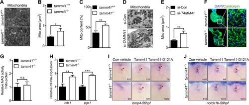
Tamm41 deficiency leads to cardiomyocytes mitochondrial defects. ac TEM images show enlarged mitochondria (lower panel) in the heart tissues of tamm41−/− embryos compared with mitochondria (upper panel) in tamm41+/+ cardiomyocytes. b and c are quantitative data for mitochondrial areas and mitochondrial contents in tamm41+/+ and tamm41−/− hearts based on TEM images respectively. d, e TEM analysis of human cardiomyocyte cell line (AC16) reveals enlarged mitochondria formation with TAMM41 deficiency. Mitochondrial area quantitation is shown in (e). f Immunofluorescence analysis of cardiolipin revealed no obvious differences in fluorescence signals between tamm41+/+ and tamm41−/− hearts. Right are the enlarged ones. Scale bar: 20 μm. g Relative CL level assessed by NAO in mitochondria isolated from 52hpf tamm41+/+ and tamm41−/− embryos. h Elevated cardiolipin synthesis-related genes (cds1 and pgs1) in tamm41−/− embryos. i, j WISH assessment of valve-related markers with tamm41, tamm41-D121A mRNA overexpression. Tamm41-D121A, which has been suggested defective in cardiolipin synthesis, harbors the same effects in restoring the heart valves of tamm41−/− embryos (third panel in i and j, red arrows) as with tamm41 overexpression (second panel in i and j, red arrows). Black horizontal lines indicate mean ± SD. Means ± SD are shown for three independent experiments. n.s, not significant, *P < 0.05; **P < 0.01; ***P < 0.001 (Student’s t-test)

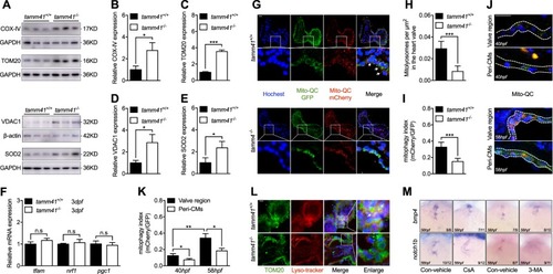
Disturbed mitophagy progression tamm41 deficient embryos. aeTamm41−/− embryos exhibit increased IMM (COX-IV), OMM (VDAC1, TOM20), and matrix (SOD2) protein levels revealed via western blot analysis. f Normally expressed mitochondrial biogenesis genes (tfam, nrf1, pgc1) in tamm41−/− embryos revealed by qPCR. gi The mito-QC was used to detect the lysosome-located mitochondria in tamm41+/+ and tamm41−/− hearts. Reduced red fluorescence intensity in tamm41−/− heart valve, which is not observed in the circulating red blood cells. For each group, ten hearts were examined. Scale bar: 20 μm. j, k mito-QC examination of mitophagy activities at 40 hpf and 58 hpf hearts. For each heart (n = 10), a ratio of mCherry to GFP mean fluorescence intensity was generated for the valve or peri-cardiomyocytes (Peri-CMs) region. Scale bar: 20 μm. l Representative images manifest the increased delivery of mitochondria (TOM20, green) to lysosome (lyso-tracker, red) in tamm41+/+ hearts (shown by white arrows), as compared with that in tamm41−/− hearts. For each group, ten hearts were examined. Scale bar: 20 μm. m Reduced heart valve genes, including bmp4 and notch1b, caused by either CsA or 3-MA addition. Black horizontal lines indicate mean ± SD. Means ± SD are shown for three independent experiments. n.s, not significant, *P < 0.05; **P < 0.01; ***P < 0.001 (Student’s t-test)

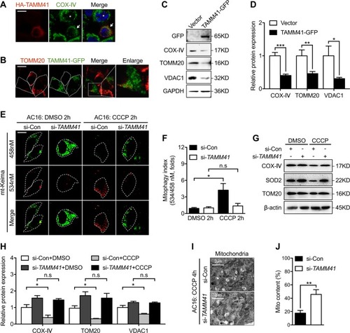
Tamm41 regulates mitophagy progression in AC16 cells. a COS7 cells with HA-TAMM41 (red) overexpression manifest fragmented mitochondrial (COX-IV; green) appearance (white arrowhead) compared with cells without HA-TAMM41 transfection (asterisk). Scale bar: 5 μm. b Immunofluorescence assay shows diminished endogenous mitochondria (TOMM20, red) in TAMM41-GFP overexpressed 293T cells. Scale bar: 5 μm. c, d TAMM41 overexpression reduced mitochondrial protein levels in AC16 cells. e, f Mitophagy activity assessed using mt-Keima in si-control or si-TAMM41 AC16 cells. Elevated levels of mitophagy were observed following CCCP treatment in control AC16 cells, which is not detected in TAMM41 deficient ones. n = 50 cells calculated per group. Scale bar: 5 μm. g, h Western blot analysis reveal compromised CCCP-induced mitochondrial membrane protein elimination ability in TAMM41-deficient cells. i, j TEM (i) and mitochondrial contents calculation (j) show that while mitochondria engulfed by autophagosome (indicated by white arrow) and disappeared after CCCP treatment in si-control AC16 cells, enlarged mitochondria remain abundant (lower panel in i) in TAMM41 deficient AC16 cells. Black horizontal lines indicate mean ± SD. Means ± SD are shown for three independent experiments. n.s, not significant, *P < 0.05; **P < 0.01; ***P < 0.001 (Student’s t-test)

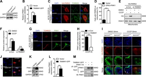
Tamm41 is required for the PARK2-PINK1 mitophagy process. a, b Elevated unstabilized PINK1 (50KD) in tamm41−/− embryos. c, d Representative images (n = 50 cells per group) show that TAMM41 silencing affected CCCP induced HA-PARK2 (red) recruitment onto mitochondria (TOMM20, green) in AC16 cells. Scale bar: 20 μm. e, f Western blot assessment of mitochondrial-fractionated PARK2 levels in transfected AC16 cells. TAMM41 deficiency inhibited PARK2 mitochondrial accumulation elicited by CCCP addition. g, h Representative images (n = 50 cells per group) show that reducing TAMM41 expression impeded CCCP triggered PINK1-GFP mitochondrial stabilization (TOMM20, red). Scale bar: 20 μm. i Representative images (n = 10 per group) reveal that CCCP treatment induced HA-PARK2 (red) translocation onto mitochondria (TOM20, green) in tamm41+/+ hearts (white arrows), but failed to do so in tamm41−/− hearts. Scale bar: 20 μm. j Enhanced HA-PARK2 recruitment onto mitochondria in TAMM41-GFP overexpressed AC16 cells (white arrowhead) compared with cell without TAMM41 overexpression (asterisk). Scale bar: 20 μm. k, l TAMM41-GFP overexpression promoted PINK1 stabilization (60KD) in the AC16 cell line by western blot. m Co-immunoprecipitation assay demonstrates the interaction between TAMM41-GFP and PINK1-V5. Black horizontal lines indicate mean ± SD. Means ± SD are shown for three independent experiments. n.s, not significant, **P < 0.01; ***P < 0.001 (Student’s t-test)

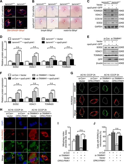
Mitochondria tethered Pink1 overexpression restores tamm41 deficiency induced heart valve abnormalities. a Representative images show the recovering effect of opa3-pink1 overexpression on 56 hpf tamm41−/− heart valve via DR-GRASP staining. Scale bar: 20 μm. bOpa3-pink1 injection at one-cell stage embryos restored heart valve markers, including bmp4 and notch1b, in tamm41−/− embryos. c, d Reduced mitochondrial membrane protein levels (fourth lane) in tamm41−/− embryos with Opa3-pink1 mRNA injection. ef OPA3-PINK1 overexpression displayed reduced mitochondrial membrane proteins in si-TAMM41 AC16 cells with CCCP addition (fourth panel). g Representative images (n = 50 per group) revealed mito-tethered PINK1 (OPA3-PINK1) overexpression restored CCCP triggered mitophagy activity in the TAMM41 deficient AC16 cell line. Scale bar: 5 μm. h Representative images (n = 50 cells per group) show the enforced OPA3-PINK1 restored HA-PARK2 translocation onto mitochondria (indicated by white arrows) in the TAMM41 deficient AC16 cell line with CCCP treatment. Scale bar: 20 μm. i, j (i) and (j) are quantitative data for (g) and (h) respectively. Black horizontal lines indicate mean ± SD. Means ± SD are shown for three independent experiments. n.s, not significant, *P < 0.05; **P < 0.01; ***P < 0.001 (Student’s t-test)

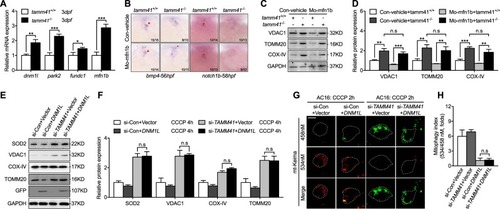
Enforced mitochondrial fission couldn’t restore heart valve malformation with tamm41 mutant embryos. a qPCR analysis of mitophagy related gene expression (dnm1l, park2, fundc1, mfn1b) in tamm41+/+ and tamm41−/− zebrafish embryos. b Representative images reveal the failure of MO-mfn1b injection to recover the abnormally expressed heart valve markers in tamm41−/− embryos (lower panels). Red arrows indicate the normally expressed heart valve genes. c, d Representative images of three independent experiments revealed that elevated mitochondrial membrane proteins in tamm41−/− embryos could not be restored with MO-mfn1b injection. e, f DNM1L-GFP overexpression failed to decrease the elevated mitochondrial membrane proteins in si-TAMM41 AC16 cells with CCCP treatment (fourth panels). g, h Representative images (n = 50 cells) show that DNM1L-GFP overexpression is incapable of restoring defective mitophagy in TAMM41 deficient AC16 cells with CCCP addition. Scale bar: 5 μm. Black horizontal lines indicate mean ± SD. Means ± SD are shown for three independent experiments. n.s, not significant, *P < 0.05; **P < 0.01; ***P < 0.001 (Student’s t-test)

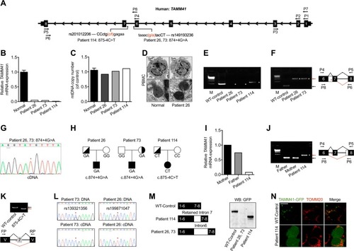
Variations of TAMM41 identified in AVSD patients. a Schematic diagram of TAMM41 transcript 005 (Ensemble ID: OTTHUMT00000339255) and the variations in Patient 26, 73, and 114 with their virtual positions in intron 6. b qPCR analysis of TAMM41 from whole blood cell extracted RNA with primers P2 and P3 (shown in a). The results show significantly reduced TAMM41 expression in the three affected children compared with six normal controls. c mtDNA copy number were evaluated by assessing the relative amounts of mitochondrial DNA and nuclear DNA (nDNA) of PBMCs from three patients and six healthy controls. MT-CO2 was used for as a marker for mtDNA and GAPDH for nDNA. d Representative TEM images showing mitochondrial morphology of PBMCs isolated from Patient 26 and one healthy control. Lower are enlarged ones showing mitochondria of the upper images. e Nest PCR with primers P1-P5 and P7-P6 (shown in a) failed to amplify full-length TAMM41 in Patient 26 and 73, and revealed an abnormally longer product in Patient 114. f Left is the DNA agarose gel electrophoresis showing that whilst normally spliced transcripts including exon 6, 7, and 8 (indicated by black arrows) amplified with primers P4 and P5 in control samples, the intron 6 retained transcript was detected via nested PCR (P4-P5 and P8-P6, indicated in a) in Patient 26 and 73. Right is the schematic drawing showing the different splicing patterns between the normal control (black dashed lines) and the patient samples (red dashed lines). g Sequencing of the PCR products in Patient 26, 73 (Fig. 1f) revealed the transcription of which from the mutant allele (indicated by red asterisks). h Transmission pattern of the TAMM41 variations identified in Patient 26, 73, and 114. i, j qPCR analysis indicating normal TAMM41 expression in sample from the father of Patient 114. The amplification products using P4-P5 and P8-P6 of Patient114 (red arrow), and her parents (black arrow) are shown on the left. Right is the schematic drawing indicating the different splicing events. k The variation (875-4C>T) in the splicing minigene reporter caused inactivation of the nearby splicing acceptor site and led to exon 7-skipping transcript generation. Lower is the schematic drawing manifesting the splicing pattern. l Exclusive expression from one allele was found in Patient 26 (rs199871047) and Patient 73 (rs139321356). The difference between genomic genotype (double peaks) and cDNA genotype (single peak) in Patient 26 and 73 implied the monoallelic expression of TAMM41. Red asterisks indicate the SNP positions. m Western blot analysis reveals that while a truncated protein caused by a premature stop codon was generated by the aberrant transcript identified in Patient 114, no protein was detected from the intron 6 inserted construct (Patient 26 and 73). n Co-immunofluorescence of mitochondrial outer membrane marker TOMM20 (red) and TAMM41-GFP showed that the truncated TAMM41, generated from Patient 114’s aberrant transcript, failed to localize onto mitochondria. Scale bar: 5 μm. V, vector. M, marker. White asterisks in e, f and j indicate transcripts generated by nest PCR. Red asterisks in f, g, j and k indicate the variation site

Acknowledgments
This image is the copyrighted work of the attributed author or publisher, and ZFIN has permission only to display this image to its users. Additional permissions should be obtained from the applicable author or publisher of the image. Full text @ Cell Death Differ.