IMAGE

Fig. 1

ID
ZDB-IMAGE-191220-4
Genes
Source
Figures for Yang et al., 2019
Image
Figure Caption

Fig. 1

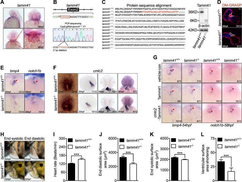
Tamm41 is indispensable for zebrafish cardiogenesis. a WISH analysis of tamm41 expression in zebrafish embryos at 36 hpf and 48 hpf. Red dotted lines delineate how tamm41 expressed. b Schematic diagram of tamm41 targeted crispr9 design, and transmittable F1 carrying 7 bp deletion in the exon2 of tamm41 was generated. c The deletion caused a frame shift and generated a premature stop codon leading to truncated Tamm41 (6KD) generation without most of the functional domains. d Representative images show DR-GRASP staining of 56 hpf tamm41+/+ and tamm41−/− hearts. While several layers of myocardium (upper panel outlined by white dashed line) form adjacent to the endocardial cushion in tamm41+/+ hearts, only a single layer of cardiomyocytes (lower panel outlined by white dashed line) is observed in tamm41−/− hearts. Scale bar: 20 μm. e Representative images show the expression of heart valve formation markers bmp4 and notch1b at the indicated times. While these markers are restrictively expressed in the AV canal region in tamm41+/+ embryos (upper panels, red arrows), they are weakly diffused throughout the whole hearts of tamm41−/− embryos (lower panels). f Representative images of WISH assay of cardiomyocyte maturation marker cmlc2 expression at the indicated times in tamm41+/+ and tamm41−/− embryos. No obvious differences were detected. White dashed lines indicate the heart morphology outlined by cmlc2 expression. g WISH analysis of bmp4 and notch1b expression with tamm41 or cmlc2-tamm41 overexpression in tamm41+/+ and tamm41−/− embryos. The abnormal bmp4 and notch1b expression patterns in tamm41−/− hearts are restored with either enforced tamm41 or cmlc2-tamm41 expression. Red arrows indicate normally restricted expression of bmp4 or notch1b in the heart valve. hl Optical Heartbeat analysis of cardiac function. Representative images showing ventricular at peak diastole and systole stage which are taken from movies of beating hearts (h). The heart rates (i), end-diastolic area (EDA) (j), and end systole area (ESA) (k) of tamm41+/+ and tamm41−/− embryos (52 hpf, n = 10 for each group) are calculated by measuring the corresponding long (black line; a) and short (red line; b) axis (EDA or ESA = a/2 × b/2 × π). Fractional area changes of the ventricle (ventricular surface area shortening) (l) are measured by (EDA-ESA)/EDA × 100). Data are presented as mean ± SD. ***P < 0.001 (Student’s t-test)

Figure Data
Acknowledgments
This image is the copyrighted work of the attributed author or publisher, and ZFIN has permission only to display this image to its users. Additional permissions should be obtained from the applicable author or publisher of the image. Full text @ Cell Death Differ.