- Title
-
Specification of sensory neurons occurs through diverse developmental programs functioning in the brain and spinal cord
- Authors
- Dyer, C., Linker, C., Graham, A., Knight, R.
- Source
- Full text @ Dev. Dyn.
|
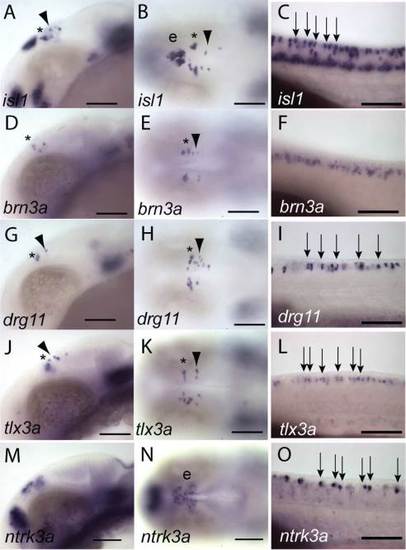
Heterogeneity of transcription factor gene expression in primary neuronal populations of the central nervous system. The expression of isl1 (A–C), brn3a (D-F), drg11 (G–I), tlx3a (J–L), and ntrk3a (trkc1, M–O) was analysed in 24-hpf embryos by in situ hybridisation. Expression of isl1, brn3a, drg11, and tlx3a was observed in MTN neurons (arrowheads), or nTPC neurons (asterisk) as well as RB neurons (arrows). In contrast, brn3a was observed in few MTN and nTPC neurons and was not observed in RB neurons, but instead was restricted to neurons at more ventral neural tube positions (D–F). We did not detect any expression of ntrk3a in nTPC or MTN neurons although we observed expression in epiphyseal neurons (e) and cells of the posterior diencephalon (M-N). In contrast, ntrk3a was expressed in a sub-population of RB neurons relative to isl1 expression (O). EXPRESSION / LABELING:
|
|
MTN neurons do not arise from neural crest and do not require sox10 function for their development. Expression (blue) of isl1 (A,C) and drg11 (B) in MTN (arrows) and nTPC neurons does not overlap with expression of foxd3 (A,B) and sox10 (C) in mesencephalic neural crest (red) in 24-hpf embryos processed by double in situ hybridisation. Similarly, there is no co-localisation of Isl1 protein (D,F) or drg11 expression (G,I) in MTN neurons with EGFP (E,F,H,I) in neural crest of 24-hpf Tg[foxd3:egfp] embryos, although there is co-localisation in the epiphysis (e). There is no apparent difference in the number of isl1 expressing MTN neurons (J,K) or RB neurons (L,M) in cls mutants (K,M) relative to wildtype (WT) siblings (J,L) at 26 hpf. Quantification of RB, MTN, and nTPC neurons in cls and WT embryos reveals no significant (NS) differences except for a difference in nTPC number (*P<0.05, error bars represent standard error of the mean, N). Scale bars = 100 µm (A–M). EXPRESSION / LABELING:
PHENOTYPE:
|
|
Notch signalling components are expressed in the midbrain at early neurogenesis stages. Lateral views of the heads of 14-hpf embryos processed by in situ hybridisation (A–V). The Notch receptors elta, deltaB, deltaC, and deltaD (A–H), the Notch ligands notch1a, notch1b, notch2, and notch3 (I–P) and Notch-regulated genes her4, hey21, and hey4 (Q-V) were assessed for expression in the dorsal midbrain where MTN neurons will arise (bracket). No specific delta gene expression was detected, but notch1b and notch2 expression was observed in the dorsal midbrain. Both hey2 and her4 also showed expression in dorsal midbrain cells. Dorsal and orthogonal views of a 24-hpf Tg[elavl3:egfp] embryo processed by fluorescent in situ hybridisation and immunohistochemistry with anti-GFP reveals that expression of deltaB did not co-localise with EGFP in MTN neurons (intersect lines, W). Scale bars = 100 µm (A–W). EXPRESSION / LABELING:
|
|
Neural crest and RB neuron development is Notch-dependent, but MTN and nTPC neurons do not require Notch signalling for their development. Embryos treated with 100 µM DAPT or DMSO from 30% epiboly were fixed at 24 hpf (A,B,E,F) or 19 hpf (C,D,G,H) and processed by in situ hybridisation with probes for isl1 (A,B,E,F) or crestin (C,D,G,H). In animals treated with DAPT, lateral aspects of the spinal cord reveal that there are many more RB neurons (arrows) relative to DMSO treated controls (A,B). Commensurate with this, there are less migrating neural crest cells (arrows) at spinal cord levels in DAPT-treated animals relative to controls (C,D). Dorsal views of the brain reveal that there are no obvious changes to nTPC (asterisk) or MTN (arrowheads) neurons in DAPT-treated embryos (E,F). However, lateral views of the head reveal that there are less migrating mesencephalic neural crest cells (arrows) in DAPT-treated animals relative to DMSO treated controls (G,H). Quantification of neurons revealed that DAPT treatment caused a significant reduction of RB neurons (**P<0.01), but no significant change (NS) to MTN or nTPC numbers. Scale bars = 100 µm (A–H). |
|
Ngn1 function is required for RB neuron development, but not for MTN development. A–C: Dorsal views of the brains of 24-hpf Tg[elavl3:egfp] embryos processed by fluorescent in situ for ngn1 and labelled with anti-GFP. Orthogonal views at the level of MTN neurons show there is no co-localisation of ngn1 and EGFP in nTPC (asterisk) or MTN neurons (arrowheads, C). D–F: A comparison of huc/elavl3 expression to GFP in the brain of a 24-hpf Tg[-8.4neurog1:GFP] transgenic larvae does not reveal co-localisation in nTPC or MTN neurons, although some epiphyseal neurons (ep) do show co-expression. Uninjected (G,I,K) or embryos injected with ngn1 morpholino (H,J,L), then fixed at 24 hpf. Lateral views of the spinal cord reveal that isl1 expression in absent from RB neurons in ngn1 morphants (G,H). A dorsal view of the spinal cord labelled with zn-12 reveals a loss of RB neurons (arrows) and reticulospinal tract axons (I,J). Dorsal views of the head reveal that isl1 expressing MTN and nTPC neurons appear to be unaffected in ngn1 morphants (K,L). M: Quantification of RB, MTN,and nTPC neurons expressing drg11 reveals that ngn1 loss of function results in a dramatic reduction of RB neurons (**P<0.01), but does not result in a significant change (NS) of MTN or nTPC neuron number. |


