- Title
-
The spinster homolog, two of hearts, is required for sphingosine 1-phosphate signaling in zebrafish
- Authors
- Osborne, N., Brand-Arzamendi, K., Ober, E.A., Jin, S.W., Verkade, H., Holtzman, N.G., Yelon, D., and Stainier, D.Y.
- Source
- Full text @ Curr. Biol.
|
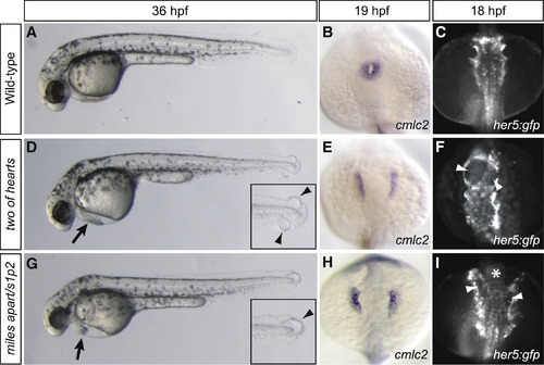
two of hearts and miles apart Mutant Phenotypes Comparison of WT (A–C), toh mutant (D–F), and mil/s1p2 mutant (G–I) embryos. (A, D, and G) Lateral brightfield images, anterior to the left, at 36 hpf show pericardial edema (arrow) and epidermal blisters (insets, arrowheads) in the tails of tohsk12 (D) and mil/s1p2m93 (G) mutants. (B, E and H) Examination of cmlc2 expression at 19 hpf shows heart-ring formation in WT embryos (B) and a failure in precardiac-mesoderm migration in tohs420 (E) and mil/s1p2m93 (H) mutant embryos. Dorsal views with anterior upwards. (C, F and I) Visualization of the anterior endoderm by Tg(-0.7her5:EGFP)ne2067 expression at 18 hpf. In embryos injected with toh (F) and mil/s1p2 (I) MOs, numerous gaps (arrowheads) appear in the endodermal sheet, which is also irregularly shaped. The most anterior region of mil/s1p2 morphants lacks GFP+ endodermal cells at the midline (asterisk). Dorsal views with anterior upwards. EXPRESSION / LABELING:
PHENOTYPE:
|
|
Isolation of the two of hearts Gene (A) Positional cloning of toh. Direction of the chromosomal walk is indicated with black arrows above the marker names. Markers used for mapping are indicated above the line representing the genomic region. The numbers of recombination events out of 2394 meioses found at each marker are indicated below the genomic region. A magnification of the critical region depicts portions of six open-reading frames (open arrows) in the identified genetic interval, including the previously cloned locus liebeskummer/reptin. The critical region contains two spinster-like genes, one of which was identified as toh (gray filled arrow) and the other of which we here name spinl3. (B) Schematic diagram of Toh proteins produced from each allele. Arrows point to the site affected in the mutant alleles. Transmembrane domains are indicated in blue, and the predicted translated intronic sequence in the s8 allele is indicated in red. Numbers also identify the individual transmembrane domains. (C) Injection of a splice blocking MO against toh into WT embryos phenocopies the toh mutations, leading to pericardial edema (arrow) and blistering in the tail (arrowhead). PHENOTYPE:
|
|
Toh Function in the YSL Is Required for Precardiac-Mesoderm Migration (A–D) toh expression at 6 (A and B) and 8 (C and D) hpf. (A) Animal-pole view with dorsal to the right, 6 hpf, showing toh expression around the margin and diffusely throughout the YSL. (B) Magnified view of the box in (A) showing toh expression around the YSL nuclei (arrowhead). (C) Lateral view with dorsal to the right, 8 hpf, showing continued toh expression in cells that have involuted, as well as in the YSL. (D) Magnified view of the box in (C) showing pronounced toh expression in the YSL (arrowhead). (E and F) Dorsal images of Tg(cmlc2:GFP)f1 embryos injected into the YSL with mock solution (E) or with 4 ng toh MO (F) and visualized at 30 hpf. Mock-injected embryos have a single heart tube (E, arrow), whereas embryos with loss of Toh function in the YSL very frequently (84%, n = 56) display cardia bifida (F; arrows). |
|
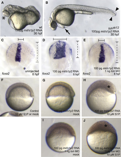
Exogenous S1P Substitutes for Toh Function in Mil/S1P2-Overexpressing Embryos (A and B) Lateral views with anterior to the left, of embryos overexpressing mil/s1p2 at 30 hpf. WT embryos injected with 100 pg of mil/s1p2 RNA (A) display a shortened body axis and cyclopia (asterisk). MZtohs8 mutants injected with 100 pg of mil/s1p2 RNA have no body-axis defects (B) but develop pericardial edema (arrow) and tail blisters (arrowheads). (C–E) Visualization of axial mesoderm and endoderm by expression of foxa2 at late gastrula stages, dorsal views with anterior upwards. WT embryos overexpressing mil/s1p2 (D) have broadened axial mesoderm (solid line) compared to uninjected embryos (C). Progression of epiboly (dashed line) is also impaired in embryos overexpressing mil/s1p2 as compared to uninjected siblings. toh MO-injected embryos overexpressing mil/s1p2 (E) are indistinguishable from uninjected siblings (C). (F–J) Lateral views of 5.5 hpf embryos injected with mock carrier solution or 10 μM S1P. Control embryos show no response to exogenous S1P (F). Embryos overexpressing Mil/S1P2 do not show more severe phenotypes when injected with mock carrier solution at 5 hpf (G). Mil/S1P2-overexpressing embryos injected with a 10 μM S1P solution at 5 hpf exhibit severe phenotypes (H), including an exaggerated thickened layer of cells at the animal pole (asterisk). Mil/S1P2-overexpressing embryos coinjected with toh MO show no phenotypes when injected with mock solution (I) and resemble controls (F). When injected with a 10 μM S1P solution, Mil/S1P2-overexpressing embryos lacking Toh function show severe defects (J), including a thickened layer of cells at the animal pole (asterisk). EXPRESSION / LABELING:
|
|
toh Is Expressed Dynamically throughout Development Images of toh expression during early development. toh mRNA is maternally provided as seen at the 8 cell stage (A, dorsal view). In a lateral view at 4 hpf, diffuse toh expression can be seen throughout the blastoderm (B). At 6 hpf (C, lateral view, dorsal to the right), toh expression is upregulated in cells that have undergone involution (arrowhead) and is also expressed in the yolk cell. At 24 hpf (D-G), toh is expressed in distinct domains. In a whole animal lateral view, anterior to the left (D), expression can be seen in the endoderm, which is adjacent to the yolk. In a blow up of the boxed region in panel D, toh expression can be seen in a distinct compartment within the somites (E, arrowhead). In dorsal, anterior (F) and dorsolateral, anterior (G) views, anterior to the left, toh expression can be seen in the heart tube. |
|
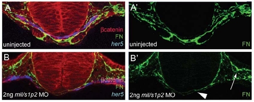
Fibronectin Expression Is Affected in mil MO-Injected Embryos Confocal micrographs of cross sections of uninjected (A and A′) and injected with 2 ng of mil MO (B and B′) Tg(-0.7her5:EGFP)ne2067 embryos (GFP false colored blue) stained for β-catenin (red) to show cellular outlines and Fibronectin (green) at 20 hpf. Control embryos (A and A′) have endodermal cells (blue) spanning the midline (blue) and show strong Fibronectin deposition (green) under the endoderm and around the migrating precardiac mesoderm. mil MO injected embryos (B and B′) show gaps in the endodermal sheet (blue) and have considerably less Fibronectin deposition (green) at the midline (arrowhead), while lateral Fibronectin deposition (arrow) appears relatively unaffected. |


