- Title
-
Zebrafish mutants with disrupted early T-cell and thymus development identified in early pressure screen
- Authors
- Trede, N.S., Ota, T., Kawasaki, H., Paw, B.H., Katz, T., Demarest, B., Hutchinson, S., Zhou, Y., Hersey, C., Zapata, A., Amemiya, C.T., and Zon, L.I.
- Source
- Full text @ Dev. Dyn.
|
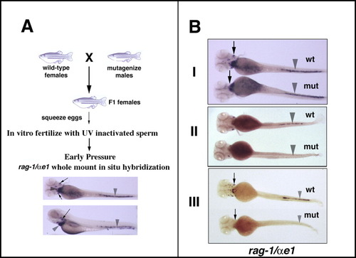
Design of screen and classes of mutants obtained. A:Schematic diagram of screen design. For details, refer to the Results and Experimental Procedure sections. Whole-mount in situ hybridization (WISH) at 5 days postfertilization (dpf) wild-type (wt) larvae with rag-1 and αe1 probes shows expression of rag-1 in bilateral thymi (arrows) and αe1 in the heart and tail (arrowhead). B:Three classes of mutants were observed in the screen. In each panel the wild-type (wt) larvae are on top, the mutant (mut) larvae are on the bottom. I. Lymphoid mutants showed normal expression of αe1 (arrowheads) but absence of rag-1 expression (arrows) compared with wt controls (top panel). II. Erythroid mutants showed normal expression of rag-1, but defective expression of αe1 (middle panel). III. Mutants showing defects in both rag-1 and αe1 expression point to a possible defect in the early hematopoietic compartment (bottom panel). |
|
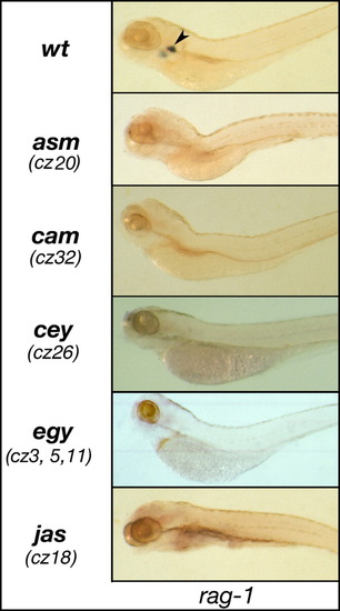
Lymphoid mutants recovered from screen. rag-1 whole-mount in situ hybridization (WISH) in 5 days postfertilization (dpf) wild-type (arrowhead, top panel) and mutant (lower panels) larvae assam (asm), chamomile (cam), ceylon (cey), and jasmine (jas). Allele designations are given below mutant names in parentheses. |
|
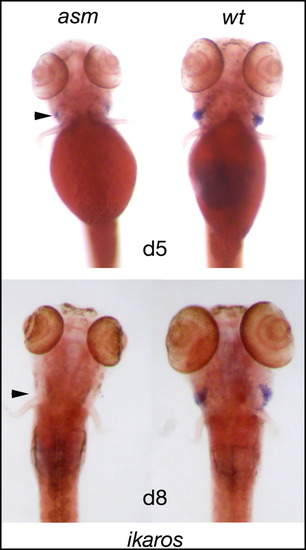
Ikaros expression in wild-type and asm mutants. Ventral view of ikaros expression in wild-type (right) and asm mutant (left) lavae at 5 dpf (top panel) and 8 days postfertilization (dpf; lower panel). ikaros expression is seen in wt larvae in the thymic area (arrowhead). In asm mutants, ikaros expression is seen at 5 dpf, but absent at 8 dpf. |
|
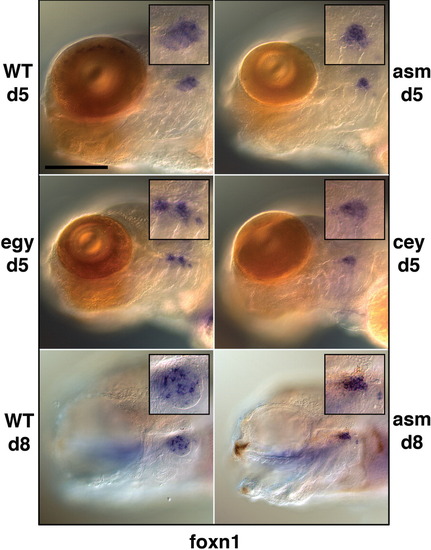
foxn1 expression in selected mutants. Lateral view of 5 days postfertilization (dpf; top four panels) and 8 dpf (bottom panels) foxn1 expression in wild-type and mutant larvae at x20 magnification. Insert shows x40 magnification of thymic signal. Scale bar = 200 μm. For better mounting and imaging, the left eye was removed from 8 dpf larvae. Note the wide dispersion of the foxn1 signal in 8 dpf wild-type larva (bottom left panel). |
|
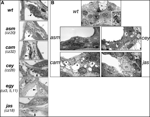
Abnormal thymic architecture in mutant larvae. A: Light microscopic examination of wild-type 7 dpf thymus shows a heterogeneous, cellular thymus (dark arrowhead). Epithelium of the otic capsule is indicated by grey arrowhead. Thymus of cey and cam mutant larvae shows reduction in thymic epithelial cell layers and empty spaces (arrows). Magnification was x100 for all panels. B:Electron microscopy of 7 dpf wt thymus (top panel) shows a heterogeneous population of cells. Arrowheads indicate basement membrane. Insert (x54,000) in right lower corner shows desmosomes (D, arrow) between thymic epithelial cells. Occasional lymphoblasts were only found in asm mutants. Empty spaces suggesting degeneration in the thymic epithelial cells were encountered in cey and cam mutants (asterisks). Magnification was x4,000 for all panels. Ch, chloride cell, a salt-producing cell occurring in the epithelium of pharyngeal cavity of many fish species; Fb, fibroblast; Lb, lymphoblast; Mu, mucine producing cell; Ph, pharyngeal epithelial cell; TEC, thymic epithelial cell. PHENOTYPE:
|
|
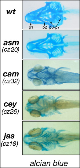
Abnormalities in pharyngeal arch architecture in mutant larvae. Architecture of pharyngeal arches by alcian blue staining shows presence of seven fully chondrified arches in 7 days postfertilization (dpf) wild-type larvae (indicated by arrows, p1 to p7). Various defects are seen in cey and cam mutants, while asm resembles wt morphology and jas has a complete set of shortened arches. PHENOTYPE:
|
|
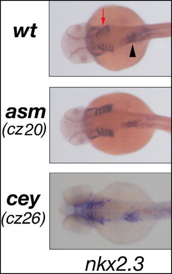
nkx2.3 expression in wt and selected mutant embryos. In dorsal view of 2 days postfertilization (dpf) wild-type (wt; top panel) and mutant (lower panels) two nkx2.3 expression domains are observed: pharyngeal pouches (red arrow) and gut tube (arrowhead). EXPRESSION / LABELING:
|

Unillustrated author statements |

