- Title
-
Endothelin 1-mediated regulation of pharyngeal bone development in zebrafish
- Authors
- Kimmel, C.B., Ullmann, B., Walker, M., Miller, C.T., and Crump, J.G.
- Source
- Full text @ Development
|
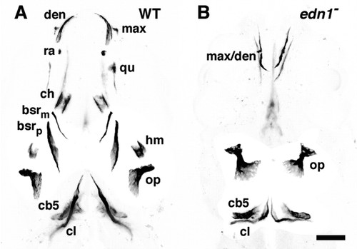
Ossifications in the young wild-type (WT) zebrafish (A) and homozygous edn1 mutant (B). Ventral views, anterior to the top, of negative images of bones fluorescently labeled with Calcein in larvae at 7-days postfertilization. Ventral bones of the pharyngeal arches are identified (Cubbage and Mabee, 1996) by their labels on the left side, and dorsal bones are labeled on the right side in both panels, all of them are present as bilateral pairs. (A) The wild type first or mandibular arch includes a dorsal and a ventral dermal bone, the maxilla (max) and dentary (den), and a dorsal and a ventral cartilage-replacement bone, the quadrate (qu) and retroarticular (ra). The second or hyoid arch includes a dorsal and two ventral dermal bones, opercle (op), and two branchiostegal rays (bsrp and bsrm), and dorsal and ventral cartilage replacement bones (very incompletely ossified at this stage), the hyomandibula (hm) and ceratohyal (ch). The most posterior arch includes a cartilage-replacement bone, ceratobranchial 5 (cb5). Overlaying ceratobranchial 5 is the cleithrum (cl), a long dermal bone connecting the posterior skull and the pectoral girdle. Two other craniofacial bones present at this stage lie deeper in the tissue and are not labeled, the parasphenoid and the endopterygoid. (B) Many of the anterior ossifications (in the first two arches) are missing in the edn1 mutant. Ceratobranchial 5 and the cleithrum are present, shortened and somewhat malformed. In the mandibular arch dermal bones (max/den) are present but severely malformed, an example of the `wicket′ phenotype discussed in the text (see also Fig. 3). In the hyoid arch the opercle is present and its joint region (upper part of the bone) is markedly expanded, a mild example of the `opercle-gain′ phenotype described in the text and other Figures. Scale bar: 100 μm. PHENOTYPE:
|
|
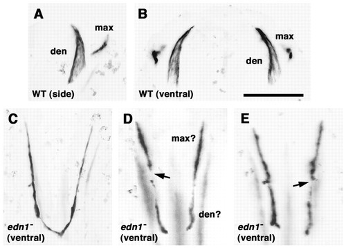
Dermal bones of the mandibular arch and their transformations in edn1 mutants. Anterior is to the top. The wild-type (WT) maxilla (max) and dentary (den) are shown in right-side view (dorsal to the right) (A) and ventral view (B). (C-E) Three examples of the AP-reversed wicket phenotype in three individual edn1 mutants, that we interpret to include the malformed bilateral rudiments of the maxillas and dentaries, completely fused (C), or incompletely fused together (D,E) (ventral views, see also Fig. 2 for orientation). The arrows in D and E indicate variably present gaps along the wicket arms. In examples not shown with only mild reduction of Edn1 (as obtained in sturgeon mutants or when edn1-MO is injected at low levels), the maxilla and dentary are frequently normally shaped, but misoriented: the orientation relates to the change in the shape of the mouth, as in edn1 mutants. Injected at higher levels, edn1-MO yields the wicket phenotype, phenocopying the edn1 mutants, and a yet more extreme phenotype in which only a small bone fragment is present (data not shown). This remaining element is usually located at an anterior position in the wall of the mouth, suggesting that it is a remnant of the maxilla. Scale bar: 100 μm. PHENOTYPE:
|
|
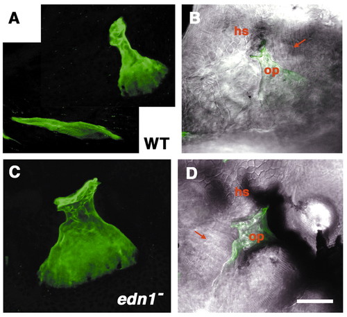
The opercle-gain phenotype in an edn1 mutant. Left-side views with dorsal to the top at 6-days postfertilization. (A,B) Wild type (WT), (C,D) mutant. (A,C) Projections of the Calcein-labeled bones from z-stacks of sections made by confocal microscopy. (B,D) The same projections are overlaid onto Nomarski differential interference contrast (DIC) images at the plane of focus of the joint made by the opercle (op) with the hyosymplectic cartilage (hs). The opercle (green) at this stage is fan-shaped in the wild type (A). It is similarly shaped but larger in the mutant (B); the joint region (upper) is thicker and the fan is expanded. The DIC views reveal that the opercle-gain joints are made with the hyosymplectic cartilage as in the wild type. In these two panels (B,D) the cells of the hyosymplectic cartilage can be recognized by their characteristic mosaic-tile shapes, and are present just above the opercle. Muscles, identifiable by their striated appearance (arrows), connect with wild type and mutant opercles. Often in the mutants, as here to the left side, ectopic muscles connect to the gain opercle. Scale bar: 50 μm. PHENOTYPE:
|
|
Two classes of opercle osteoblast clusters, expanded and reduced, are present in she mutants. Labeling with the zns5 monoclonal antibody in larvae at 88 hours postfertilization. Left-side views with dorsal to the top. The antibody labels both osteoblasts and nerve axons prominently, and muscles lightly. (A) wild type (WT). Osteoblasts envelop the opercle (arrow) and branchiostegal ray (asterisk). The dark lines are nerves. Delicately labeled muscles (at the top) connect separately to the opercle joint region (dilator operculi muscle, left) and blade region (adductor operculi, right). (B) A she mutant with a presumed mild opercle-loss phenotype. The branchiostegal ray osteoblast cluster is missing. Two clusters are present (arrows) that seem to correspond to the opercle joint region (upper, identified by its position and its connection with the dilator operculi muscle) and a diminished opercle `fan′ region (lower). A confocal z-series reveals no osteoblasts connect between these two clusters. (C) A she mutant with presumed opercle gain. The opercle osteoblast cluster is expanded (arrow), particularly at the joint region. The branchiostegal ray cluster is either missing or possibly is present but fused with the opercle cluster. Muscle connections are approximately normal. Scale bar: 50 μm. PHENOTYPE:
|
|
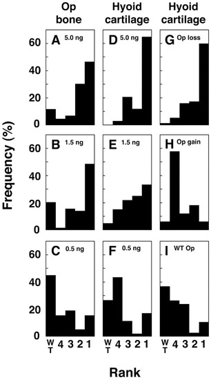
Opercle (A-C) and cartilage (D-F) phenotypes depend on the level of Edn1 reduction in edn1-MO-injected larvae, and co-vary in individual hyoid arches (G-I). The MO was injected at 5 ng, 1.5 ng and 0.5 ng. (A-F) Resulting sets of opercle and hyoid cartilage phenotypes, scored independently on both sides of individual larvae, and ranked according to severity. Facial phenotypes (not shown) are as expected (Miller and Kimmel, 2001). The highest levels of edn1-MO generally produce the more severe phenotype, resembling edn1 mutants. Milder facial phenotypes, resembling hoo- were observed at the lowest levels. The data are shown as percentages; the number of arches scored (left and right, considered separately) ranged from 33 to 75 for each panel, from a total of 73 larvae (146 sides). (A-C) Opercle ranks are: 1, missing; 2, reduced; 3, mildly expanded (Fig. 8B-F); 4, markedly expanded (Fig. 8G-K); wild type (WT), normal. (D-I) Hyoid cartilage phenotypes were scored according to degree of reduction (we do not observe completely missing cartilages or cartilage expansions), reflecting the spectrum previously shown [Fig. 9 of Kimmel et al. (Kimmel et al., 1998)]: 1 and 2: relatively severe reductions as in typical severe (1) and mild (2) edn1 mutants; 3, intermediate loss, as in typical she mutants; 4, mild loss, as in a typical stu mutants; WT, wild type. (G-I) The cartilage ranks shown in animals grouped (independently of the amount of injected MO) by their opercle phenotypes in the same (left or right) hyoid arches. Opercle ranks 1 and 2 (in A) together constitute the opercle-loss group; ranks 3 and 4 together constitute the opercle-gain group, and ranks 3 and 4 together constitute the opercle-gain group. We used Chi-square analysis (data not shown) to test and reject (P<0.001) the null hypothesis that severities of cartilage and bone phenotypes vary independently from one another in individual arches. MO, morpholino. PHENOTYPE:
|
|
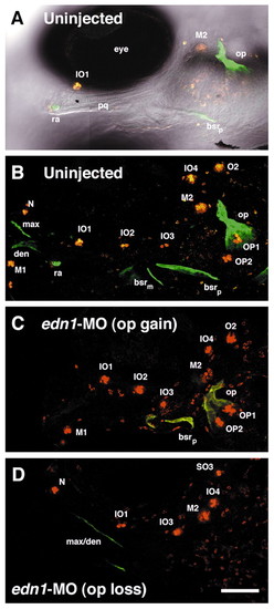
Correlated expressivities of opercle and lateral line neuromast phenotypes in edn1-MO-injected larvae (C,D), compared with an uninjected larva (A,B). Left-side views of live larvae, with dorsal to the top and anterior to the left at 6-days post fertilization. (B-D) Confocal z-series projections. (A) A single optical section from the image stack in B combining DIC with fluorescence for orientation. The bones are labeled with Calcein (green), and the neuromasts with DASPEI (orange-red). Neuromasts were identified (Raible and Kruse, 2000) and are labeled with upper case letters; other structures are labeled in lower case. (B) The normal neuromast distribution in the uninjected control. (C) An edn1-MO-injected larva expressing the opercle-gain phenotype, the branchiostegal ray (bsr) is also malformed. The complete set of neuromasts is present, however IO3 is displaced ventrally and M1 is displaced posteriorly. (D) An edn1-MO-injected larva expressing the opercle-loss phenotype. Both the opercle and branchiostegal ray are missing, and the mandibular dermal bones (max/den) resemble the condition in Fig. 3E. The neuromast phenotype in this particular larva is severe; losses include M1, IO2, O2 and the two opercular neuromasts OP1 and OP2. Scale bar: 100 μm. MO, morpholino. PHENOTYPE:
|
|
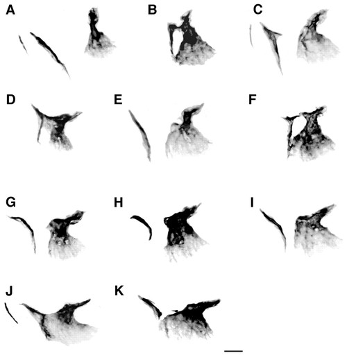
Phenotypic series of opercle-gain and branchiostegal phenotypes in larvae with mild reductions of Edn1. Left-side views (confocal projections, Calcein labeling) with dorsal to the top at 6 or 7 days postfertilization. (A) Wild type (WT). (B-K) Mutants and MO-injected larvae arranged according to the extent of the increase in size of the opercle. (B,F) she mutants. (C) edn1-MO injected at 5 ng. (E,H,I,K) edn1-MO injected at 1.5 ng. (D,G,J) stu mutants. Two branchiostegal rays are present in (A) and (C), and apparently also in (J), in which the larger dorsal one is deformed and fused with the opercle by a thin but continuous sheet of bone. We interpret (K) as a similar prominent fusion between the disrupted branchiostegal ray and the opercle. (B,D,F) Examples of `walking stick′ phenotypes, showing fusions at the joint regions of the bones. (C) The branchiostegal ray is deformed with a spur appearing to correspond to an expanded joint region, also present in the walking stick examples. The fusions seem to occur all along the series (i.e. in examples with either a mild or severe opercle gain), whereas the curvature of the branchiostegal ray roughly increases along the series with the most extreme case in H. Scale bar: 50 μm. PHENOTYPE:
|
|
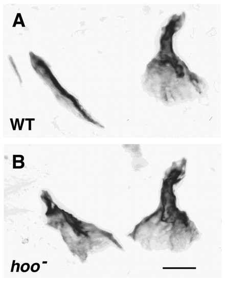
Putative homeotic transformation of the dorsal branchiostegal ray towards the opercle in a hoover mutant. (A) A wild-type (WT) hoob631 sibling, showing the opercle and two branchiostegal rays, the more anterior one much smaller. (B) A hoo mutant. The opercle (right) is approximately normal in size and shape, but the bone at the usual position of the dorsal branchiostegal ray (left) has the form of an opercle. Its proximal end (upper) is sculptured into a distinctive joint region, rather than just ending bluntly as in A. Here DIC imaging (not shown; as in Fig. 4) reveals that the transformed branchiostegal ray makes a prominent joint with the underlying ceratohyal cartilage. Its distal region, normally blade-shaped, is expanded into an opercle-like fan. The other (more anterior and smaller) branchiostegal ray is missing. Scale bar: 50 μm. PHENOTYPE:
|
|
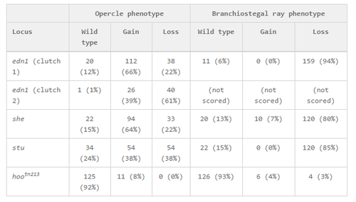
Opercle and branchiostegal ray phenotypes in anterior arch mutants. Homozygous mutants were obtained by crossing heterozygotes that were scored by facial phenotypes and separated from phenotypic wild-type siblings before fixation and bone staining with Calcein. The wild-type phenotype indicates that the element (opercle or branchiostegal ray) appears normal in shape and size. The gain phenotype means any significant expansion (see text). The loss class includes reduction in size, as well as other cases where the element is missing altogether. The two edn1 examples (separate experiments involving crosses between different individual fish) are quantitatively different, indicating that quantitative comparisons between clutches must be interpreted with caution. Similarly, in other experiments, stu mutants had higher fractions of opercle gain phenotypes, comparable with she mutants in the table. PHENOTYPE:
|
|
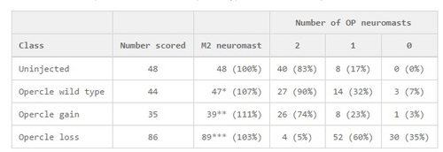
Correlations between opercle and neuromast phenotypes in edn1-MO-injected larvae. We scored presence or absence of neuromasts in the pharyngeal region (see their labeling in Fig. 7), and (on the same sides of the same larvae) the bone phenotypes. For the table, classes were assigned by phenotype of the opercle, irrespective of amount of injected edn 1-MO. The frequency distributions for the bone and cartilage phenotypes from the same set of larvae are shown in Fig. 6. As the phenotype varies between the two sides of a single larva, each side of each larva was scored separately, and ′Number scored′ refer to sides scored. We used Chi-square analysis (data not shown) to test and reject (P<0.001) the null hypothesis that severities of cartilage and OP neuromast phenotypes vary independently from one another in individual arches. ↵* In three cases, the M2 neuromast was duplicated, resulting in a total of 47. ↵** In one case, no M2 neuromasts were present; in five cases the M2 neuromast was duplicated, resulting in a grand total of 39. ↵*** In seven cases, no M2 neuromasts were present; in ten cases the M2 neuromast was duplicated, resulting in a grand total of 89. PHENOTYPE:
|

