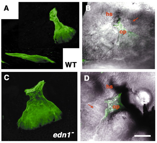
The opercle-gain phenotype in an edn1 mutant. Left-side views with dorsal to the top at 6-days postfertilization. (A,B) Wild type (WT), (C,D) mutant. (A,C) Projections of the Calcein-labeled bones from z-stacks of sections made by confocal microscopy. (B,D) The same projections are overlaid onto Nomarski differential interference contrast (DIC) images at the plane of focus of the joint made by the opercle (op) with the hyosymplectic cartilage (hs). The opercle (green) at this stage is fan-shaped in the wild type (A). It is similarly shaped but larger in the mutant (B); the joint region (upper) is thicker and the fan is expanded. The DIC views reveal that the opercle-gain joints are made with the hyosymplectic cartilage as in the wild type. In these two panels (B,D) the cells of the hyosymplectic cartilage can be recognized by their characteristic mosaic-tile shapes, and are present just above the opercle. Muscles, identifiable by their striated appearance (arrows), connect with wild type and mutant opercles. Often in the mutants, as here to the left side, ectopic muscles connect to the gain opercle. Scale bar: 50 μm.
|
