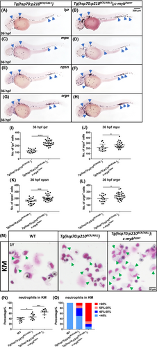
Fig. 3 Exogenous c-myb activation can mimic CML progression in the zebrafish CML model. (A–H) WISH showed increase of lyz (A,B), mpx (C,D), npsn (E,F), and srgn (G,H) expression in Tg(hsp70:p210BCR/ABL1);c-mybhyper at 36 hpf compared with Tg(hsp70:p210BCR/ABL1). Blue arrowheads indicate lyz+, mpx+, srgn+ and npsn+ neutrophils in each row. (I–L) Quantification of numbers of 36 hpf lyz+ (I) (Tg(hsp70:p210BCR/ABL1), n = 16; Tg(hsp70:p210BCR/ABL1);c-mybhyper, n = 30), mpx+ (J) (Tg(hsp70:p210BCR/ABL1), n = 15; Tg(hsp70:p210BCR/ABL1);c-mybhyper, n = 25), npsn+ (K) (Tg(hsp70:p210BCR/ABL1), n = 22; Tg(hsp70:p210BCR/ABL1);c-mybhyper, n = 31), srgn+ (L) (Tg(hsp70:p210BCR/ABL1), n = 18; Tg(hsp70:p210BCR/ABL1);c-mybhyper, n = 32) cells. (O) May-Grünwald Giemsa staining of kidney marrow (KM) blood cells that were obtained from WT, Tg(hsp70:p210BCR/ABL1), and Tg(hsp70:p210BCR/ABL1);c-mybhyper adult zebrafish. Green arrowheads indicated neutrophils. After staining, 1000 cells were randomly chosen for further calculation. (N) The proportion of neutrophils in white blood cells in whole kidney marrow (WT, n = 10; Tg(hsp70:p210BCR/ABL1), n = 10; Tg(hsp70:p210BCR/ABL1);c-mybhyper, n = 10). (O) High proportions of neutrophils presented more in Tg(hsp70:p210BCR/ABL1);c-mybhyper than in Tg(hsp70:p210BCR/ABL1). Scale bars, 200 μm (A–H) and 50 μm (M).
Image
Figure Caption
Figure Data
Acknowledgments
This image is the copyrighted work of the attributed author or publisher, and
ZFIN has permission only to display this image to its users.
Additional permissions should be obtained from the applicable author or publisher of the image.
Full text @ Animal Model Exp Med

