IMAGE

Fig. 3.

ID
ZDB-IMAGE-210904-24
Source
Figures for Lee et al., 2021
Image
Figure Caption

Fig. 3.

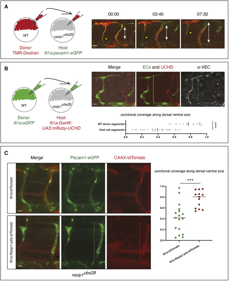
Autonomous requirement of Rasip1 during ISV formation. (A) Schematic showing transplantation of TMR-labeled donor wild-type (WT) cells into rasip1ubs28 mutant hosts with both donor and host cells expressing Pecam1-EGFP in endothelial cells. Time-lapse images of wild-type cells (red) in rasip1ubs28 mutant hosts showing that wild-type cells elongate and maintain junctional contacts (Movie 6). Double-headed arrows indicate the extent of the ISV between DA and DLAV. (B) Schematic showing transplantation of EGFP-labeled wild-type donor cells [Tg(flia:EGFP)y1] into rasip1 mutant host embryos [Tg(flia:Galff)ubs3; (UAS:UCHD-mRuby2)ubs20]. Immunofluorescence analysis of VE-cadherin in embryos at 32 hpf. Transplanted wild-type cells elongate and form multicellular tubes. Graph shows quantification of junctional coverage along the dorsal-ventral axis (ratio between the accumulated length of junctions to the length of the ISV) (analyzed WT donor segment n=20, host cell segment n=23). (C) Rescue by transient endothelial-specific rasip1 expression. Endothelial expression was achieved by DNA microinjection using a fli1a:Rasip1-p2a-tdTomato-CAAX construct or fli1a:tdTomato-CAAX as a control. Graph shows quantification of junctional coverage along the dorsal-ventral axis (analyzed ISVs in rasip1ubs28 mutant background n=17, rasip1 over-expression n=13). Scale bars: 20 μm. Analyzed by unpaired two-tailed Mann–Whitney test (***P<0.0001 in B, ***P<0.001 in C); error bars indicate s.d.

Acknowledgments
This image is the copyrighted work of the attributed author or publisher, and ZFIN has permission only to display this image to its users. Additional permissions should be obtained from the applicable author or publisher of the image. Full text @ Development