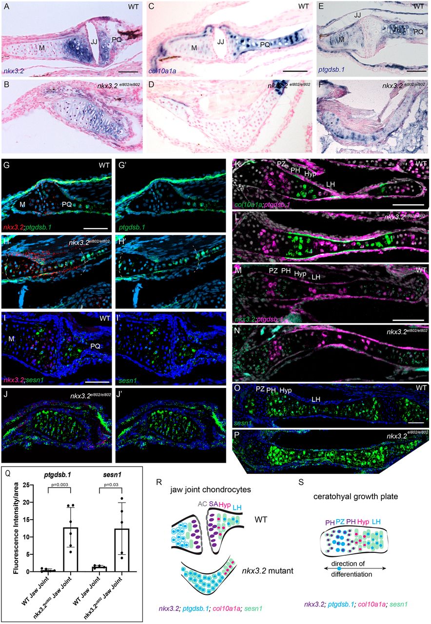
Fig. 6 Increased expression of ptgdsb.1 and sesn1 across the fused jaw joint of nkx3.2 mutant zebrafish. (A,B) Colorimetric in situ hybridization demonstrates nkx3.2 expression in the sub-articular zone of the jaw joint and across the fused jaw joint in nkx3.2el802 mutants at 21 dpf. (C-F) Colorimetric in situ hybridization shows expansion of ptgdsb.1 but not col10a1a across the fused jaw joint region of nkx3.2 mutants. (G-J′) RNAScope in situ hybridization shows ectopic expression of ptgdsb.1 (G-H′) and sesn1 (I-J′) across the fused jaw joint region of mutants (identified by nkx3.2 expression in red). DAPI labels nuclei in blue. Dashed lines in H,H′ indicate fused jaw joint region. (K-P) RNAscope in situ hybridizations show expression of col10a1a, ptgdsb.1, nkx3.2 and sesn1 in the growth plates of the ceratohyal. (Q) Quantification of fluorescence intensity at the jaw joint demonstrates upregulation of ptgdsb.1 and sesn1 in nkx3.2 mutants. Individual animals are plotted (dots) with mean±s.d. (R,S) Model of chondrocyte zones at the wild-type and fused mutant jaw joint (R) and within the bidirectional ceratohyal growth plate (S). AC, articular surface; Hyp, hypertrophic; JJ, jaw joint; LH, late hypertrophic; M, Meckel's cartilage; PH, pre-hypertrophic; PQ, palatoquadrate; PZ, proliferative zone; SA, sub-articular zone; WT, wild type. Scale bars: 50 µm.
Image
Figure Caption
Figure Data
Acknowledgments
This image is the copyrighted work of the attributed author or publisher, and
ZFIN has permission only to display this image to its users.
Additional permissions should be obtained from the applicable author or publisher of the image.
Full text @ Development

