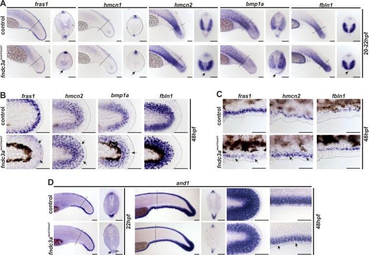
Normal development of the ventral median fin fold is altered in fndc3awue1/wue1 mutants. (A) Investigation of fras1, hmcn1, hmcn2, bmp1a, and fbln1 expression in the tail bud 20–22 hpf indicated loss of mesenchymal cells in the median fin fold in fndc3awue1/wue1 mutants (arrows; fras1: 9/32; hmcn1: 11/26; hmcn2: 24/43; bmp1a: 22/45; fbln1: 16/32). (B) Expression of mesenchymal markers in posterior fins of 48 hpf old embryos was slightly changed following loss of fndc3a (fras1: 10/24; hmcn2: 12/25; bmp1a: 8/21; fbln1: 9/24). (C) Ventral fin folds in 48 hpf old embryos did not show loss of mesenchymal markers after fndc3awue1/wue1 mutation. (D) Expression of and1, an essential marker for actinotrichia formation, in fndc3awue1/wue1 in the ventral fin fold was partly lost in 22 hpf embryos (12/25), but recovered in 48 hpf old embryos to a reduced expression level (control n = 16; weaker expression in fndc3awue1/wue1 embryos n = 10/18). Dashed lines in (A,D) indicate planes of shown sections. Dashed lines in (B,C) indicate fin boundaries. Scale bars: 50 µm
This image is the copyrighted work of the attributed author or publisher, and
ZFIN has permission only to display this image to its users.
Additional permissions should be obtained from the applicable author or publisher of the image.
Full text @ Sci. Rep.
Your Input Welcome
Thank you for submitting comments. Your input has been emailed to ZFIN curators who may contact you if
additional information is required.
Oops. Something went wrong. Please try again later.