IMAGE

Fig. 2.

ID
ZDB-IMAGE-190723-894
Genes
Source
Figures for Cho et al., 2019
Image
Figure Caption

Fig. 2.

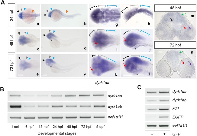
Zebrafish dyrk1aa and dyrk1ab are expressed in the developing brain during embryogenesis and detected in endothelial cells of the vasculature. (A) WISH showed that dyrk1aa was expressed in the forebrain (black arrowheads, Aa-Af; black brackets, Ag-Al), the midbrain (gray arrowheads, Aa-Af; gray brackets, Ag-Al), the hindbrain (blue arrowheads, Aa-Af; blue brackets, Ag-Al) at 24, 48 and 72 hpf, and the spinal cord (orange arrowheads, Aa and Ab) at 24 hpf. It was also detected in the heart (black asterisks, Ad,Af,Aj,Al) and in the retina (red arrows, Ai-Al) at 48 and 72 hpf. Am and An are sectioned images of WISH embryos showing the expression of dyrk1aa in the tectum (green arrows), tegmentum (black arrow), the ganglionic cell layer (purple arrows) and the inner nuclear layer of the retina (red arrows) at 48 hpf and 72 hpf. See Fig. S3 for WISH of dyrk1ab. (B) Semi-quantitative RT-PCR analysis of dyrk1aa and dyrk1ab mRNA expression in whole embryos at indicated developmental stages. dyrk1aa and dyrk1ab expression was observed at the one-cell stage followed by diminishing at 6 hpf and resuming after 24 hpf. (C) Semi-quantitative RT-PCR analysis using endothelial cells isolated by FACS for sorting GFP-positive and -negative cells of Tg(kdrl:EGFP) embryos at 48 hpf revealed that dyrk1aa and dyrk1ab mRNAs were expressed in GFP-positive endothelial cells as well as in GFP-negative embryonic cells. Scale bars: 200 µm in Aa-Al; 50 µm in Am,An.

Figure Data
Acknowledgments
This image is the copyrighted work of the attributed author or publisher, and ZFIN has permission only to display this image to its users. Additional permissions should be obtained from the applicable author or publisher of the image. Full text @ Dis. Model. Mech.