Fig. 2
Fig. 2
Transformation of the Upper Jaw in Nr2f Mutants
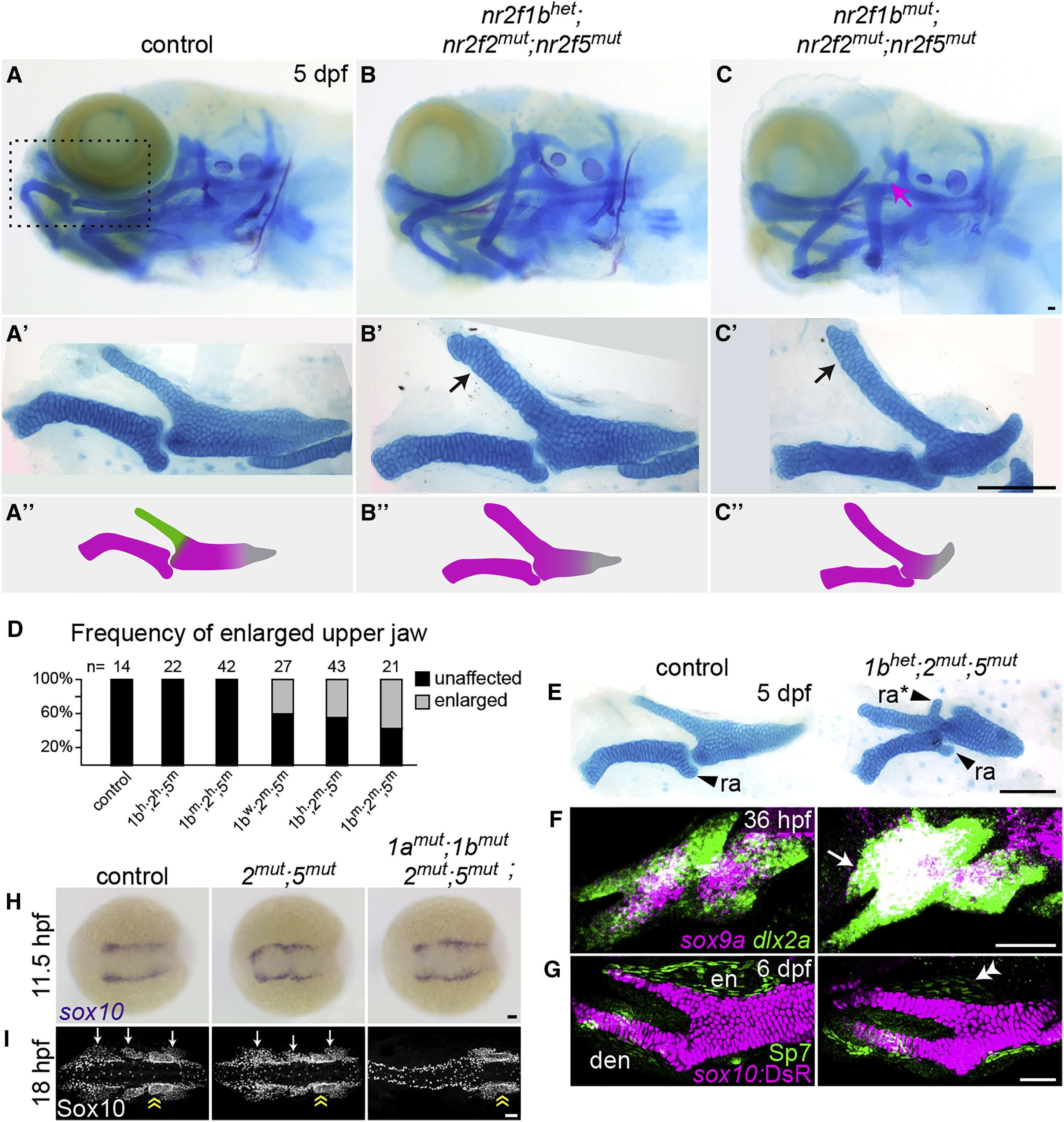
(A–C) Larval heads stained for cartilage (Alcian blue) and bone (Alizarin red). Boxed area in (A) represents the approximate region of the dissected upper- and lower-jaw cartilages in A′–C′. In mutants (B′–C′), the pterygoid process (Ptp, green in A″) of the palatoquadrate is thickened (black arrows) to resemble Meckel's (magenta in A″). Skeletal structures derived from the dorsal hyoid arch are reduced in the triple mutant (magenta arrow in C).
(D) Frequency of the thickened Ptp phenotype across different nr2f1b; nr2f2; nr2f5 genotypes.
(E) Some nr2f2; nr2f5 mutants form an ectopic process (ra∗) that resembles the retroarticular process (ra) of Meckel's.
(F) Fluorescent in situ images show expansion of sox9a expression into the mutant maxillary prominence (white arrow; dlx2a labels all arch NCCs).
(G) Formation of a larger cartilage (sox10:DsRed+, magenta) in the mutant is accompanied by a reduction in the number of Sp7+ osteoblasts (green) in the neighboring dermal entopterygoid bone (en; white double arrowhead). Mutants have normal numbers of Sp7+ osteoblasts in the lower-jaw dentary bone (den).
(H) sox10+ NCCs are specified at the neural plate border in all Nr2f mutants (n = 18 nr2f1aany; nr2f1bany; nr2f2−/−; nr2f5−/−; n = 2 quadruple). The nr2f2; nr2f5 double mutant shown is also nr2f1a+−-; nr2f1b−/−.
(I) Three streams of Sox10+ NCCs (white arrows) are evident in all mutant combinations (n = 13 nr2f1aany; nr2f1bany; nr2f2−/−; nr2f5−/−) except the quadruple mutant (n = 1). Sox10+ otic cells (yellow double arrow) are indicated for reference.
Scale bars: (C and E) 100 μm; (F–I) 50 μm. Also see Figure S2 and Table S4 for mutation details and skeletal phenotypes of single and other combinatorial mutants.
Reprinted from Developmental Cell, 44(3), Barske, L., Rataud, P., Behizad, K., Del Rio, L., Cox, S.G., Crump, J.G., Essential Role of Nr2f Nuclear Receptors in Patterning the Vertebrate Upper Jaw, 337-347.e5, Copyright (2018) with permission from Elsevier. Full text @ Dev. Cell

