Fig. 5
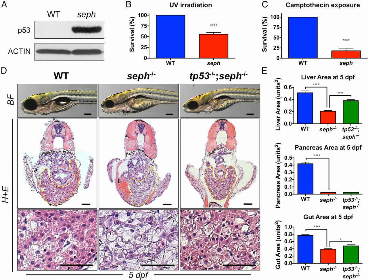
seph mutants are sensitive to DNA damage. (A) Immunoblot analysis of p53 expression in WT and seph mutant larvae at 5 dpf. (B) Survival of WT and seph mutant larvae 2 dpe to UV irradiation (200 J/m2, 7 dpf). n = 7. ****P < 0.0001. (C) Survival of WT and seph mutant larvae 2 dpe to camptothecin (1 µM). n = 10. ****P < 0.0001. (D) Morphological and histological analysis of WT, seph-/- and tp53-/-;seph-/- mutant larvae at 5 dpf. The yellow dashed region represents the liver. The region of histology showing the larval liver is shown in the zoomed images. [Scale bars: 200 µm (brightfield; BF) and 50 µm (histology; H+E).] (E) Quantitative analysis of liver, pancreas, and gut area in WT, seph-/-, and tp53-/-;seph-/- mutant larvae at 5 dpf. n > 8. *P < 0.05; ****P < 0.0001.

