Fig. 4
Fig. 4
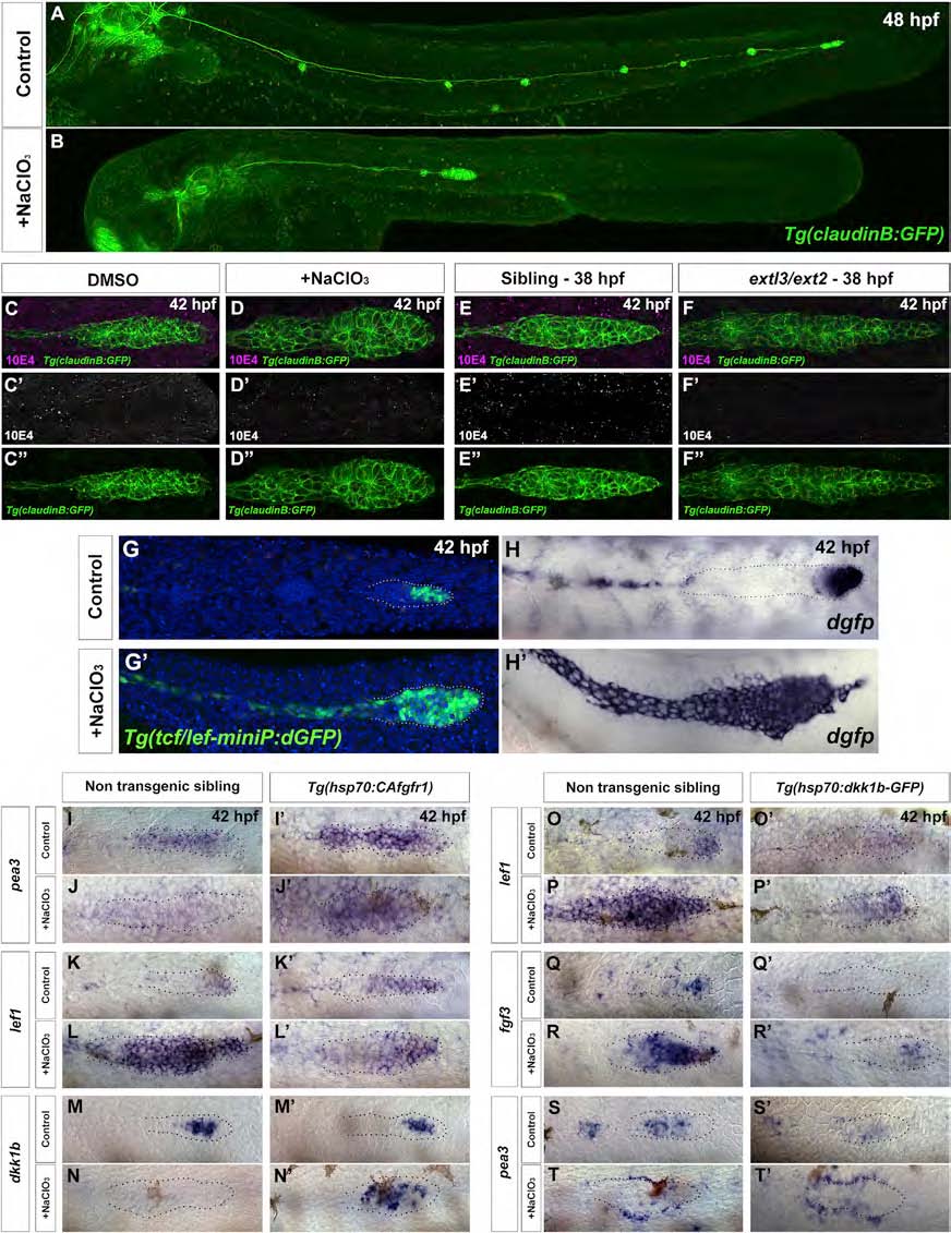
NaClO3 Treatment Phenocopies extl3/ext2 Mutants, Causing Loss of Fgf Signaling and Resulting in Expansion of the Wnt Domain
(A and B) In NaClO3-treated embryos, the prim stalls.
(C-D′′) NaClO3-treated embryos show downregulation of HS levels by 10E4 antibody staining.
(E-F′′) extl3/ext2 mutants show a similar loss of HS.
(G-H′) NaClO3-treated Wnt reporter line embryos show expansion of the Wnt domain in the prim, as evidenced by destabilized GFP (dGFP) expression and dgfp in situ hybridization.
(I-J′) The Fgf target pea3 is restored in NaClO3-treated prim after CAfgfr1 induction.
(K-L′) Restored Fgf signaling restricts expression of the Wnt target lef1 back to the leading region.
(M-N′) The restored restriction correlates with the activation of dkk1b after Fgf pathway activation.
(O-T′) Wnt is restricted after NaClO3 treatment by inducing Dkk1b expression in the Tg(hsp70:dkk1b-GFP) line. In the absence of HS, Dkk1b induction is sufficient to restrict the Wnt targets lef1 and fgf3 back to the leading region (O-P′ and Q-R′), but not to restore Fgf signaling in the trailing region of the prim (S-T′). pea3 is still only expressed in cells surrounding the prim (T′).
See also Figure S4, Table S1, and Movies S5 and S7.

