Image
Figure Caption
Fig. 2
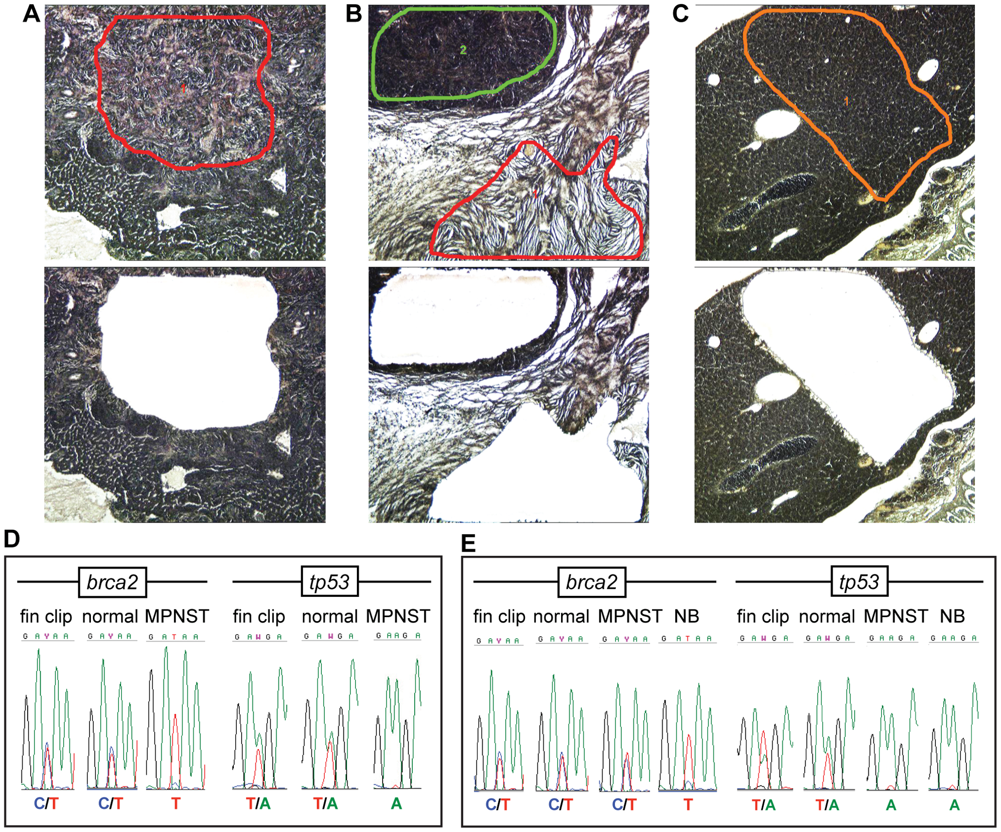
Malignant zebrafish tumors frequently develop LOH for brca2 and/or tp53.
(A–C), Before (upper panels) and after (lower panels) images of LCM-guided sample collection from an MPNST (A), an MPNST and a nephroblastoma (B), and a normal liver (C). Regions of sample collection are outlined in color. (D) MPNST from a brca2+/m;tp53+/m zebrafish shows loss of the brca2 and tp53 wildtype alleles. (E) MPNST and nephroblastoma from a brca2+/m;tp53+/m zebrafish show disparate LOH profiles. LOH, loss of heterozygosity; MPNST, malignant peripheral nerve sheath tumor; NB, nephroblastoma.
Figure Data
Acknowledgments
This image is the copyrighted work of the attributed author or publisher, and
ZFIN has permission only to display this image to its users.
Additional permissions should be obtained from the applicable author or publisher of the image.
Full text @ PLoS One

