Fig. 2
Fig. 2
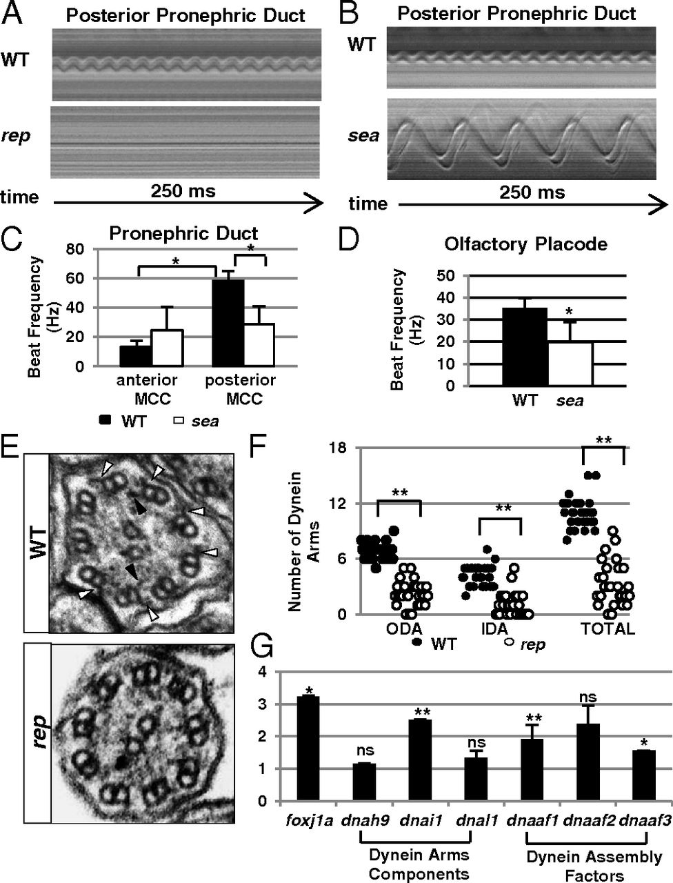
Reptin is essential for cilia motility. (A and B) Kymographs showing the rhythmic beating of a multicilia bundle in the posterior pronephric duct in a wild-type (WT) embryo and paralyzed cilia in a reptinhi2394 mutant (rep), or a slower beating cilium in a seahorsehi3308 mutant (sea) at 3 dpf. (C) Cilia beating frequency in wild-type (black bar) and seahorsehi3308 (white bar) pronephros at 3 dpf. Data are represented as mean + SD, from eight replicates. *P < 0.05. (D) Cilia beating frequency in the olfactory placode of wild-type (black bar, WT) and seahorsehi3308 mutant (white bar, sea) embryos at 3 dpf. Data are represented as mean + SD, from five replicates. *P < 0.05. (E) Electron micrographs of cross-sections of cilia in WT and reptinhi2394 mutants (rep) at 5 dpf. Outer and inner dynein arms are pointed out by white and black arrowheads, respectively. (F) Number of outer and inner dynein arms in WT and reptinhi2394 mutants (rep) per section at 5 dpf. Data are represented as mean + SD. **P < 0.01. (G) Expression levels of multiple factors involved in dynein arm formation as shown by quantitative PCR (qPCR). Y axis shows the fold changes in reptinhi2394 mutants. Data are represented as mean + SD, from three replicates. *P < 0.05, **P < 0.01. NS, nonsignificant.

